


 Foundational characteristics of cancer include proliferation, angiogenesis, migration, evasion of apoptosis, and cellular immortality. Find key markers for these cellular processes and antibodies to detect them.
 The SUMOplot™ Analysis Program predicts and scores sumoylation sites in your protein. SUMOylation is a post-translational modification involved in various cellular processes, such as nuclear-cytosolic transport, transcriptional regulation, apoptosis, protein stability, response to stress, and progression through the cell cycle.
 The Autophagy Receptor Motif Plotter predicts and scores autophagy receptor binding sites in your protein. Identifying proteins connected to this pathway is critical to understanding the role of autophagy in physiological as well as pathological processes such as development, differentiation, neurodegenerative diseases, stress, infection, and cancer.
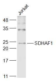
SDHAF1 Polyclonal Antibody
Purified Rabbit Polyclonal Antibody (Pab)
- SPECIFICATION
- CITATIONS
- PROTOCOLS
- BACKGROUND

Application
| WB, IHC-P, IHC-F, IF, ICC |
|---|---|
| Primary Accession | A6NFY7 |
| Reactivity | Rat, Pig, Dog, Bovine |
| Host | Rabbit |
| Clonality | Polyclonal |
| Calculated MW | 21 KDa |
| Physical State | Liquid |
| Immunogen | KLH conjugated synthetic peptide derived from human SDHAF1 |
| Epitope Specificity | 1-100/115 |
| Isotype | IgG |
| Purity | affinity purified by Protein A |
| Buffer | 0.01M TBS (pH7.4) with 1% BSA, 0.02% Proclin300 and 50% Glycerol. |
| SUBCELLULAR LOCATION | Mitochondrion matrix. |
| SIMILARITY | Belongs to the complex I LYR family. SDHAF1 subfamily. |
| DISEASE | Mitochondrial complex II deficiency |
| Important Note | This product as supplied is intended for research use only, not for use in human, therapeutic or diagnostic applications. |
| Background Descriptions | The succinate dehydrogenase (SDH) complex (or complex II) of the mitochondrial respiratory chain is composed of 4 individual subunits. The protein encoded by this gene resides in the mitochondria, and is essential for SDH assembly, but does not physically associate with the complex in vivo. Mutations in this gene are associated with SDH-defective infantile leukoencephalopathy (mitochondrial complex II deficiency).[provided by RefSeq, Mar 2010] |
| Gene ID | 644096 |
|---|---|
| Other Names | Succinate dehydrogenase assembly factor 1, mitochondrial, SDH assembly factor 1, SDHAF1, LYR motif-containing protein 8, SDHAF1 {ECO:0000303|PubMed:19465911, ECO:0000312|HGNC:HGNC:33867} |
| Target/Specificity | Ubiquitously expressed. |
| Dilution | WB=1:500-2000,IHC-P=1:100-500,IHC-F=1:100-500,ICC=1:100-500,IF=1:100-500 |
| Storage | Store at -20 ℃ for one year. Avoid repeated freeze/thaw cycles. When reconstituted in sterile pH 7.4 0.01M PBS or diluent of antibody the antibody is stable for at least two weeks at 2-4 ℃. |
| Name | SDHAF1 {ECO:0000303|PubMed:19465911, ECO:0000312|HGNC:HGNC:33867} |
|---|---|
| Function | Plays an essential role in the assembly of succinate dehydrogenase (SDH), an enzyme complex (also referred to as respiratory complex II) that is a component of both the tricarboxylic acid (TCA) cycle and the mitochondrial electron transport chain, and which couples the oxidation of succinate to fumarate with the reduction of ubiquinone (coenzyme Q) to ubiquinol (PubMed:19465911, PubMed:24954417). Promotes maturation of the iron-sulfur protein subunit SDHB of the SDH catalytic dimer, protecting it from the deleterious effects of oxidants (PubMed:24954417). May act together with SDHAF3 (PubMed:24954417). Contributes to iron-sulfur cluster incorporation into SDHB by binding to SDHB and recruiting the iron-sulfur transfer complex formed by HSC20, HSPA9 and ISCU through direct binding to HSC20 (PubMed:26749241). |
| Cellular Location | Mitochondrion matrix |
| Tissue Location | Ubiquitously expressed. |
Citations (0)

Thousands of laboratories across the world have published research that depended on the performance of antibodies from Abcepta to advance their research. Check out links to articles that cite our products in major peer-reviewed journals, organized by research category.
Submit your citation using an Abcepta antibody to
info@abcepta.com, and receive a free "I Love Antibodies" mug.
info@abcepta.com, and receive a free "I Love Antibodies" mug.

Application Protocols
Provided below are standard protocols that you may find useful for product applications.
Abcepta welcomes feedback from its customers.
If you have used an Abcepta product and would like to share how it has performed, please click on the "Submit Review" button and provide the requested information. Our staff will examine and post your review and contact you if needed.
If you have any additional inquiries please email technical services at tech@abcepta.com.
$ 385.00
Cat# AP57594
Ordering Information
United States
AlbaniaAustraliaAustriaBelgiumBosnia & HerzegovinaBrazilBulgariaCanadaCentral AmericaChinaCroatiaCyprusCzech RepublicDenmarkEstoniaFinlandFranceGermanyGreeceHong KongHungaryIcelandIndiaIndonesiaIrelandIsraelItalyJapanLatviaLithuaniaLuxembourgMacedoniaMalaysiaMaltaMexicoNetherlandsNew ZealandNorwayPakistanPolandPortugalRomaniaSerbiaSingaporeSlovakiaSloveniaSouth AfricaSouth KoreaSpainSwedenSwitzerlandTaiwanTurkeyUnited KingdomUnited StatesVietnamWorldwideOthers
USA Headquarters
(888) 735-7227 / (858) 622-0099 or (858) 875-1900
Other Products
Shipping Information
Domestic orders (in stock items)
Shipped out the same day. Orders placed after 1 PM (PST) will ship out the next business day.
International orders
Contact your local distributors
