


 Foundational characteristics of cancer include proliferation, angiogenesis, migration, evasion of apoptosis, and cellular immortality. Find key markers for these cellular processes and antibodies to detect them.
 The SUMOplot™ Analysis Program predicts and scores sumoylation sites in your protein. SUMOylation is a post-translational modification involved in various cellular processes, such as nuclear-cytosolic transport, transcriptional regulation, apoptosis, protein stability, response to stress, and progression through the cell cycle.
 The Autophagy Receptor Motif Plotter predicts and scores autophagy receptor binding sites in your protein. Identifying proteins connected to this pathway is critical to understanding the role of autophagy in physiological as well as pathological processes such as development, differentiation, neurodegenerative diseases, stress, infection, and cancer.
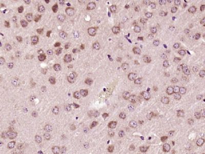
SH3BP1 Polyclonal Antibody
Purified Rabbit Polyclonal Antibody (Pab)
- SPECIFICATION
- CITATIONS
- PROTOCOLS
- BACKGROUND

Application
| IHC-P, IHC-F, IF, ICC, E |
|---|---|
| Primary Accession | Q9Y3L3 |
| Reactivity | Rat, Pig, Dog, Bovine |
| Host | Rabbit |
| Clonality | Polyclonal |
| Calculated MW | 76 KDa |
| Physical State | Liquid |
| Immunogen | KLH conjugated synthetic peptide derived from human SH3BP1 |
| Epitope Specificity | 101-200/701 |
| Isotype | IgG |
| Purity | affinity purified by Protein A |
| Buffer | 0.01M TBS (pH7.4) with 1% BSA, 0.02% Proclin300 and 50% Glycerol. |
| SIMILARITY | Contains 1 BAR domain. Contains 1 Rho-GAP domain. |
| Important Note | This product as supplied is intended for research use only, not for use in human, therapeutic or diagnostic applications. |
| Gene ID | 23616 |
|---|---|
| Other Names | SH3 domain-binding protein 1 {ECO:0000312|HGNC:HGNC:10824}, SH3BP1 (HGNC:10824) |
| Dilution | IHC-P=1:100-500,IHC-F=1:100-500,ICC=1:100-500,IF=1:100-500,ELISA=1:5000-10000 |
| Storage | Store at -20 ℃ for one year. Avoid repeated freeze/thaw cycles. When reconstituted in sterile pH 7.4 0.01M PBS or diluent of antibody the antibody is stable for at least two weeks at 2-4 ℃. |
| Name | SH3BP1 (HGNC:10824) |
|---|---|
| Function | GTPase activating protein (GAP) which specifically converts GTP-bound Rho-type GTPases including RAC1 and CDC42 in their inactive GDP-bound form. By specifically inactivating RAC1 at the leading edge of migrating cells, it regulates the spatiotemporal organization of cell protrusions which is important for proper cell migration (PubMed:21658605). Also negatively regulates CDC42 in the process of actin remodeling and the formation of epithelial cell junctions (PubMed:22891260). Through its GAP activity toward RAC1 and/or CDC42 plays a specific role in phagocytosis of large particles. Specifically recruited by a PI3 kinase/PI3K-dependent mechanism to sites of large particles engagement, inactivates RAC1 and/or CDC42 allowing the reorganization of the underlying actin cytoskeleton required for engulfment (PubMed:26465210). It also plays a role in angiogenesis and the process of repulsive guidance as part of a semaphorin-plexin signaling pathway. Following the binding of PLXND1 to extracellular SEMA3E it dissociates from PLXND1 and inactivates RAC1, inducing the intracellular reorganization of the actin cytoskeleton and the collapse of cells (PubMed:24841563). |
| Cellular Location | Cell projection. Cell junction, tight junction. Cell junction, adherens junction. Cell projection, phagocytic cup. Nucleus Cytoplasm, cytosol. Note=Localizes at the leading edge of migrating cells (PubMed:21658605, PubMed:24841563) Accumulation at forming phagocytic cups is PI3 kinase/PI3K-dependent and is specific for sites of large particles engagement and their phosphatidylinositol 3,4,5-triphosphate membrane content (PubMed:26465210). |
Research Areas
Citations (0)

Thousands of laboratories across the world have published research that depended on the performance of antibodies from Abcepta to advance their research. Check out links to articles that cite our products in major peer-reviewed journals, organized by research category.
Submit your citation using an Abcepta antibody to
info@abcepta.com, and receive a free "I Love Antibodies" mug.
info@abcepta.com, and receive a free "I Love Antibodies" mug.

Application Protocols
Provided below are standard protocols that you may find useful for product applications.
Abcepta welcomes feedback from its customers.
If you have used an Abcepta product and would like to share how it has performed, please click on the "Submit Review" button and provide the requested information. Our staff will examine and post your review and contact you if needed.
If you have any additional inquiries please email technical services at tech@abcepta.com.
$ 385.00
Cat# AP57650
Ordering Information
United States
AlbaniaAustraliaAustriaBelgiumBosnia & HerzegovinaBrazilBulgariaCanadaCentral AmericaChinaCroatiaCyprusCzech RepublicDenmarkEstoniaFinlandFranceGermanyGreeceHong KongHungaryIcelandIndiaIndonesiaIrelandIsraelItalyJapanLatviaLithuaniaLuxembourgMacedoniaMalaysiaMaltaMexicoNetherlandsNew ZealandNorwayPakistanPolandPortugalRomaniaSerbiaSingaporeSlovakiaSloveniaSouth AfricaSouth KoreaSpainSwedenSwitzerlandTaiwanTurkeyUnited KingdomUnited StatesVietnamWorldwideOthers
USA Headquarters
(888) 735-7227 / (858) 622-0099 or (858) 875-1900
Other Products
Shipping Information
Domestic orders (in stock items)
Shipped out the same day. Orders placed after 1 PM (PST) will ship out the next business day.
International orders
Contact your local distributors
