


 Foundational characteristics of cancer include proliferation, angiogenesis, migration, evasion of apoptosis, and cellular immortality. Find key markers for these cellular processes and antibodies to detect them.
 The SUMOplot™ Analysis Program predicts and scores sumoylation sites in your protein. SUMOylation is a post-translational modification involved in various cellular processes, such as nuclear-cytosolic transport, transcriptional regulation, apoptosis, protein stability, response to stress, and progression through the cell cycle.
 The Autophagy Receptor Motif Plotter predicts and scores autophagy receptor binding sites in your protein. Identifying proteins connected to this pathway is critical to understanding the role of autophagy in physiological as well as pathological processes such as development, differentiation, neurodegenerative diseases, stress, infection, and cancer.
MAP1S Antibody (Center)
Affinity Purified Rabbit Polyclonal Antibody (Pab)
- SPECIFICATION
- CITATIONS: 1
- PROTOCOLS
- BACKGROUND

Application
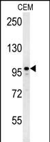
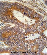
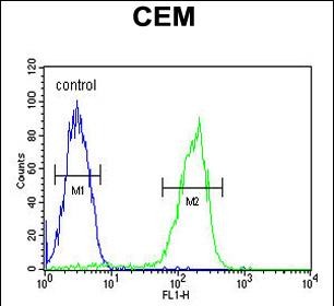
| FC, WB, IHC-P, E |
|---|---|
| Primary Accession | Q66K74 |
| Other Accession | NP_060644.4 |
| Reactivity | Human |
| Host | Rabbit |
| Clonality | Polyclonal |
| Isotype | Rabbit IgG |
| Calculated MW | 112211 Da |
| Antigen Region | 493-520 aa |
| Gene ID | 55201 |
|---|---|
| Other Names | Microtubule-associated protein 1S, MAP-1S, BPY2-interacting protein 1, Microtubule-associated protein 8, Variable charge Y chromosome 2-interacting protein 1, VCY2-interacting protein 1, VCY2IP-1, MAP1S heavy chain, MAP1S light chain, MAP1S, BPY2IP1, C19orf5, MAP8, VCY2IP1 |
| Target/Specificity | This MAP1S antibody is generated from rabbits immunized with a KLH conjugated synthetic peptide between 493-520 amino acids from the Central region of human MAP1S. |
| Dilution | FC~~1:10~50 WB~~1:1000 IHC-P~~1:50~100 E~~Use at an assay dependent concentration. |
| Format | Purified polyclonal antibody supplied in PBS with 0.09% (W/V) sodium azide. This antibody is purified through a protein A column, followed by peptide affinity purification. |
| Storage | Maintain refrigerated at 2-8°C for up to 2 weeks. For long term storage store at -20°C in small aliquots to prevent freeze-thaw cycles. |
| Precautions | MAP1S Antibody (Center) is for research use only and not for use in diagnostic or therapeutic procedures. |
| Name | MAP1S |
|---|---|
| Synonyms | BPY2IP1, C19orf5, MAP8, VCY2IP1 |
| Function | Microtubule-associated protein that mediates aggregation of mitochondria resulting in cell death and genomic destruction (MAGD). Plays a role in anchoring the microtubule organizing center to the centrosomes. Binds to DNA. Plays a role in apoptosis. Involved in the formation of microtubule bundles (By similarity). |
| Cellular Location | Nucleus. Cytoplasm, cytosol. Cytoplasm, cytoskeleton. Cytoplasm, cytoskeleton, spindle. Note=Detected in filopodia-like protrusions and synapses (By similarity). Detected in perinuclear punctate network corresponding to mitochondrial aggregates and in the nucleus in cells exhibiting apoptosis. Associated specifically with microtubules stabilized by paclitaxel and colocalizes with RASSF1 isoform A. In interphase cells, shows a diffuse cytoplasmic staining with partial localization to the microtubules. During the different stages of mitosis detected at the spindle microtubules. |
| Tissue Location | Expressed in neurons (at protein level). Expressed in spermatocytes, spermatids and spermatozoa. Expressed in the cerebral cortex. Highly expressed in testis. Moderately expressed in the brain, colon, heart, kidney, liver, lung, placenta, small intestine, spleen and stomach. Weakly expressed in muscle. |

Provided below are standard protocols that you may find useful for product applications.
Background
Microtubule-associated protein that mediates aggregation of mitochondria resulting in cell death and genomic destruction (MAGD). Plays a role in anchoring the microtubule-organizing center to the centrosomes. Binds to DNA. Plays a role in apoptosis. Involved in the formation of microtubule bundles (By similarity).
References
Beausoleil, S.A., et al. Proc. Natl. Acad. Sci. U.S.A. 101(33):12130-12135(2004)
Wong, E.Y., et al. Biol. Reprod. 70(3):775-784(2004)
Liu, L., et al. In Vitro Cell. Dev. Biol. Anim. 38(10):582-594(2002)
Liu, L., et al. Genomics 79(1):124-136(2002)
If you have used an Abcepta product and would like to share how it has performed, please click on the "Submit Review" button and provide the requested information. Our staff will examine and post your review and contact you if needed.
If you have any additional inquiries please email technical services at tech@abcepta.com.
Ordering Information
Other Products
Shipping Information
