


 Foundational characteristics of cancer include proliferation, angiogenesis, migration, evasion of apoptosis, and cellular immortality. Find key markers for these cellular processes and antibodies to detect them.
 The SUMOplot™ Analysis Program predicts and scores sumoylation sites in your protein. SUMOylation is a post-translational modification involved in various cellular processes, such as nuclear-cytosolic transport, transcriptional regulation, apoptosis, protein stability, response to stress, and progression through the cell cycle.
 The Autophagy Receptor Motif Plotter predicts and scores autophagy receptor binding sites in your protein. Identifying proteins connected to this pathway is critical to understanding the role of autophagy in physiological as well as pathological processes such as development, differentiation, neurodegenerative diseases, stress, infection, and cancer.
> home > Products > Primary Antibodies > Antibody Collections > Pancreas positive > PTPRS Polyclonal Antibody
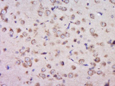
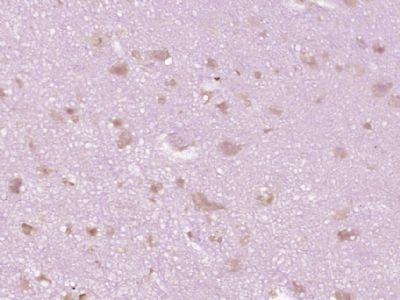
PTPRS Polyclonal Antibody
Purified Rabbit Polyclonal Antibody (Pab)
- SPECIFICATION
- CITATIONS
- PROTOCOLS
- BACKGROUND

Application
| IHC-P, IHC-F, IF, ICC, E |
|---|---|
| Primary Accession | Q13332 |
| Reactivity | Rat |
| Host | Rabbit |
| Clonality | Polyclonal |
| Calculated MW | 214 KDa |
| Physical State | Liquid |
| Immunogen | KLH conjugated synthetic peptide derived from human PTPRS |
| Epitope Specificity | 1001-1100/1948 |
| Isotype | IgG |
| Purity | affinity purified by Protein A |
| Buffer | Preservative: 0.02% Proclin300, Constituents: 1% BSA, 0.01M PBS, pH7.4. |
| SUBCELLULAR LOCATION | Membrane; Single-pass type I membrane protein. |
| SIMILARITY | Belongs to the protein-tyrosine phosphatase family. Receptor class 2A subfamily. Contains 8 fibronectin type-III domains. Contains 3 Ig-like C2-type (immunoglobulin-like) domains. Contains 2 tyrosine-protein phosphatase domains. |
| SUBUNIT | Interacts with PPFIA1, PPFIA2 and PPFIA3. |
| Post-translational modifications | A cleavage occurs, separating the extracellular domain from the transmembrane segment. This process called 'ectodomain shedding' is thought to be involved in receptor desensitization, signal transduction and/or membrane localization. |
| Important Note | This product as supplied is intended for research use only, not for use in human, therapeutic or diagnostic applications. |
| Background Descriptions | Protein tyrosine phosphatases, or PTPs, are type I transmembrane proteins, membrane associated proteins or proteins localized in nuclei. Examples of transmembrane PTPs are LAR, PTP alpha, PTP beta, PTP gamma, PTP delta, PTP epsilon, PTP zeta, PTP thet, PTP upsilon and PTPs. Transmembrane PTPsigma play diverse roles during development and in adult tissues. Immunodepletion studies have suggested LAR to be a regulator of insulin receptor phosphorylation. PTP alpha activity is increased twofold in response to phorbol ester stimulation, resulting in serine phosphorylation either directly or indirectly by members of the PKC family. Overexpression of v-H-Ras and Neu, but not Myc or Int2, in mammary tumors has been shown to induce PTPe expression. An alternative splicing event leads to a nervous tissue-specific chondroitin sulfate proteoglycan called phosphacan, which represents the amino terminal portion of PTP omega. PTP thet and PTP?share a conserved amino terminal 160 amino acid MAM domain which facilitates homophilic binding. PTP upsilon localizes to points of cell contact and may be involved in regulating the assembly and disassembly of cadherin/catenin complexes in vivo. PTPsigma contains an extracellular region, a single transmembrane segment and two tandem intracytoplasmic catalytic domains, and thus represents a receptor-type PTP. PTPsigma may also be involved in the molecular control of adult nerve repair. Four alternatively spliced transcript variants, which encode distinct proteins, have been reported. |
| Gene ID | 5802 |
|---|---|
| Other Names | Receptor-type tyrosine-protein phosphatase S, R-PTP-S, 3.1.3.48, Receptor-type tyrosine-protein phosphatase sigma, R-PTP-sigma, PTPRS |
| Target/Specificity | Detected in all tissues tested except for placenta and liver. |
| Dilution | IHC-P=1:100-500,IHC-F=1:100-500,ICC=1:100-500,IF=1:100-500,ELISA=1:5000-10000 |
| Format | 0.01M TBS(pH7.4), 0.09% (W/V) sodium azide and 50% Glyce |
| Storage | Store at -20 ℃ for one year. Avoid repeated freeze/thaw cycles. When reconstituted in sterile pH 7.4 0.01M PBS or diluent of antibody the antibody is stable for at least two weeks at 2-4 ℃. |
| Name | PTPRS |
|---|---|
| Function | Cell surface receptor that binds to glycosaminoglycans, including chondroitin sulfate proteoglycans and heparan sulfate proteoglycan (PubMed:21454754). Binding to chondroitin sulfate and heparan sulfate proteoglycans has opposite effects on PTPRS oligomerization and regulation of neurite outgrowth. Contributes to the inhibition of neurite and axonal outgrowth by chondroitin sulfate proteoglycans, also after nerve transection. Plays a role in stimulating neurite outgrowth in response to the heparan sulfate proteoglycan GPC2. Required for normal brain development, especially for normal development of the pituitary gland and the olfactory bulb. Functions as a tyrosine phosphatase (PubMed:8524829). Mediates dephosphorylation of NTRK1, NTRK2 and NTRK3 (By similarity). Plays a role in down-regulation of signaling cascades that lead to the activation of Akt and MAP kinases (By similarity). Down-regulates TLR9- mediated activation of NF-kappa-B, as well as production of TNF, interferon alpha and interferon beta (PubMed:26231120). |
| Cellular Location | Cell membrane; Single-pass type I membrane protein. Cell projection, axon {ECO:0000250|UniProtKB:B0V2N1}. Perikaryon {ECO:0000250|UniProtKB:B0V2N1}. Cytoplasmic vesicle, secretory vesicle, synaptic vesicle membrane {ECO:0000250|UniProtKB:Q64605}. Synapse, synaptosome {ECO:0000250|UniProtKB:Q64605}. Postsynaptic density {ECO:0000250|UniProtKB:Q64605}. Cell projection, neuron projection {ECO:0000250|UniProtKB:B0V2N1}. Cell projection, growth cone {ECO:0000250|UniProtKB:B0V2N1}. Note=Is rapidly internalized when dendritic cells are stimulated with the TLR9 ligand cytidine-phosphate- guanosine (CpG) (PubMed:26231120). Detected in a punctate pattern along neurites and axon growth cones (By similarity) {ECO:0000250|UniProtKB:B0V2N1, ECO:0000269|PubMed:26231120} |
| Tissue Location | Detected in peripheral blood plasmacytoid dendritic cells (at protein level) (PubMed:26231120). Detected in all tissues tested except for placenta and liver (PubMed:8524829, PubMed:8992885) Detected in peripheral blood plasmacytoid dendritic cells (PubMed:26231120). |
Research Areas
Citations (0)

Thousands of laboratories across the world have published research that depended on the performance of antibodies from Abcepta to advance their research. Check out links to articles that cite our products in major peer-reviewed journals, organized by research category.
Submit your citation using an Abcepta antibody to
info@abcepta.com, and receive a free "I Love Antibodies" mug.
info@abcepta.com, and receive a free "I Love Antibodies" mug.

Application Protocols
Provided below are standard protocols that you may find useful for product applications.
Abcepta welcomes feedback from its customers.
If you have used an Abcepta product and would like to share how it has performed, please click on the "Submit Review" button and provide the requested information. Our staff will examine and post your review and contact you if needed.
If you have any additional inquiries please email technical services at tech@abcepta.com.
$ 385.00
Cat# AP57795
Ordering Information
United States
AlbaniaAustraliaAustriaBelgiumBosnia & HerzegovinaBrazilBulgariaCanadaCentral AmericaChinaCroatiaCyprusCzech RepublicDenmarkEstoniaFinlandFranceGermanyGreeceHong KongHungaryIcelandIndiaIndonesiaIrelandIsraelItalyJapanLatviaLithuaniaLuxembourgMacedoniaMalaysiaMaltaMexicoNetherlandsNew ZealandNorwayPakistanPolandPortugalRomaniaSerbiaSingaporeSlovakiaSloveniaSouth AfricaSouth KoreaSpainSwedenSwitzerlandTaiwanTurkeyUnited KingdomUnited StatesVietnamWorldwideOthers
USA Headquarters
(888) 735-7227 / (858) 622-0099 or (858) 875-1900
Shipping Information
Domestic orders (in stock items)
Shipped out the same day. Orders placed after 1 PM (PST) will ship out the next business day.
International orders
Contact your local distributors
