


 Foundational characteristics of cancer include proliferation, angiogenesis, migration, evasion of apoptosis, and cellular immortality. Find key markers for these cellular processes and antibodies to detect them.
 The SUMOplot™ Analysis Program predicts and scores sumoylation sites in your protein. SUMOylation is a post-translational modification involved in various cellular processes, such as nuclear-cytosolic transport, transcriptional regulation, apoptosis, protein stability, response to stress, and progression through the cell cycle.
 The Autophagy Receptor Motif Plotter predicts and scores autophagy receptor binding sites in your protein. Identifying proteins connected to this pathway is critical to understanding the role of autophagy in physiological as well as pathological processes such as development, differentiation, neurodegenerative diseases, stress, infection, and cancer.
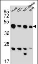
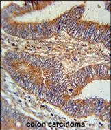
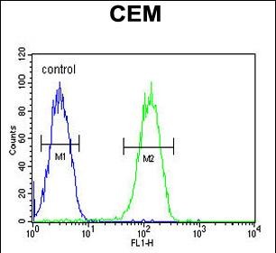
BIN2 Antibody (N-term)
Affinity Purified Rabbit Polyclonal Antibody (Pab)
- SPECIFICATION
- CITATIONS
- PROTOCOLS
- BACKGROUND

Application
| FC, IHC-P, WB, E |
|---|---|
| Primary Accession | Q9UBW5 |
| Other Accession | NP_057377.2 |
| Reactivity | Human |
| Host | Rabbit |
| Clonality | Polyclonal |
| Isotype | Rabbit IgG |
| Calculated MW | 61874 Da |
| Antigen Region | 14-42 aa |
| Gene ID | 51411 |
|---|---|
| Other Names | Bridging integrator 2, Breast cancer-associated protein 1, BIN2, BRAP1 |
| Target/Specificity | This BIN2 antibody is generated from rabbits immunized with a KLH conjugated synthetic peptide between 14-42 amino acids from the N-terminal region of human BIN2. |
| Dilution | FC~~1:10~50 IHC-P~~1:50~100 WB~~1:1000 E~~Use at an assay dependent concentration. |
| Format | Purified polyclonal antibody supplied in PBS with 0.09% (W/V) sodium azide. This antibody is purified through a protein A column, followed by peptide affinity purification. |
| Storage | Maintain refrigerated at 2-8°C for up to 2 weeks. For long term storage store at -20°C in small aliquots to prevent freeze-thaw cycles. |
| Precautions | BIN2 Antibody (N-term) is for research use only and not for use in diagnostic or therapeutic procedures. |
| Name | BIN2 |
|---|---|
| Synonyms | BRAP1 |
| Function | Promotes cell motility and migration, probably via its interaction with the cell membrane and with podosome proteins that mediate interaction with the cytoskeleton. Modulates membrane curvature and mediates membrane tubulation. Plays a role in podosome formation. Inhibits phagocytosis. |
| Cellular Location | Cytoplasm. Cell projection, podosome membrane; Peripheral membrane protein; Cytoplasmic side. Cytoplasm, cell cortex. Cell projection, phagocytic cup. Note=Associates with membranes enriched in phosphoinositides. Detected in the actin-rich cell cortex at the leading edge of migrating cells. Detected at podosomes, at an actin- rich ring-like structure. |
| Tissue Location | Detected in natural killer cells (at protein level). Highest level expression seen in spleen and peripheral blood leukocytes and is also expressed at high levels in thymus, colon and placenta, suggesting preferential expression in hematopoietic tissues |

Thousands of laboratories across the world have published research that depended on the performance of antibodies from Abcepta to advance their research. Check out links to articles that cite our products in major peer-reviewed journals, organized by research category.
info@abcepta.com, and receive a free "I Love Antibodies" mug.

Provided below are standard protocols that you may find useful for product applications.
References
Scherer, S.E., et al. Nature 440(7082):346-351(2006)
Ge, K., et al. Genomics 67(2):210-220(2000)
If you have used an Abcepta product and would like to share how it has performed, please click on the "Submit Review" button and provide the requested information. Our staff will examine and post your review and contact you if needed.
If you have any additional inquiries please email technical services at tech@abcepta.com.
Ordering Information
Other Products
Shipping Information
