


 Foundational characteristics of cancer include proliferation, angiogenesis, migration, evasion of apoptosis, and cellular immortality. Find key markers for these cellular processes and antibodies to detect them.
 The SUMOplot™ Analysis Program predicts and scores sumoylation sites in your protein. SUMOylation is a post-translational modification involved in various cellular processes, such as nuclear-cytosolic transport, transcriptional regulation, apoptosis, protein stability, response to stress, and progression through the cell cycle.
 The Autophagy Receptor Motif Plotter predicts and scores autophagy receptor binding sites in your protein. Identifying proteins connected to this pathway is critical to understanding the role of autophagy in physiological as well as pathological processes such as development, differentiation, neurodegenerative diseases, stress, infection, and cancer.
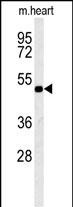
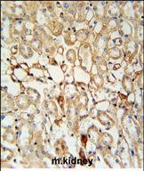
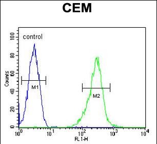
KCNV1 Antibody (N-term)
Affinity Purified Rabbit Polyclonal Antibody (Pab)
- SPECIFICATION
- CITATIONS
- PROTOCOLS
- BACKGROUND

Application
| IHC-P, FC, WB, E |
|---|---|
| Primary Accession | Q6PIU1 |
| Other Accession | P97557, Q8BZN2, Q9GKU7, Q0P583, NP_055194.1 |
| Reactivity | Human, Mouse |
| Predicted | Bovine, Monkey, Rat |
| Host | Rabbit |
| Clonality | Polyclonal |
| Isotype | Rabbit IgG |
| Calculated MW | 56304 Da |
| Antigen Region | 137-166 aa |
| Gene ID | 27012 |
|---|---|
| Other Names | Potassium voltage-gated channel subfamily V member 1, Neuronal potassium channel alpha subunit HNKA, Voltage-gated potassium channel subunit Kv81, KCNV1 |
| Target/Specificity | This KCNV1 antibody is generated from rabbits immunized with a KLH conjugated synthetic peptide between 137-166 amino acids from the N-terminal region of human KCNV1. |
| Dilution | IHC-P~~1:50~100 FC~~1:10~50 WB~~1:1000 E~~Use at an assay dependent concentration. |
| Format | Purified polyclonal antibody supplied in PBS with 0.09% (W/V) sodium azide. This antibody is purified through a protein A column, followed by peptide affinity purification. |
| Storage | Maintain refrigerated at 2-8°C for up to 2 weeks. For long term storage store at -20°C in small aliquots to prevent freeze-thaw cycles. |
| Precautions | KCNV1 Antibody (N-term) is for research use only and not for use in diagnostic or therapeutic procedures. |
| Name | KCNV1 |
|---|---|
| Function | Potassium channel subunit that does not form functional channels by itself. Modulates KCNB1 and KCNB2 channel activity by shifting the threshold for inactivation to more negative values and by slowing the rate of inactivation. Can down-regulate the channel activity of KCNB1, KCNB2, KCNC4 and KCND1, possibly by trapping them in intracellular membranes. |
| Cellular Location | Cell membrane; Multi-pass membrane protein. Note=Has to be associated with another potassium channel subunit to get inserted in the plasma membrane. Remains intracellular in the absence of KCNB2 |
| Tissue Location | Detected in brain.. |

Thousands of laboratories across the world have published research that depended on the performance of antibodies from Abcepta to advance their research. Check out links to articles that cite our products in major peer-reviewed journals, organized by research category.
info@abcepta.com, and receive a free "I Love Antibodies" mug.

Provided below are standard protocols that you may find useful for product applications.
Background
Voltage-gated potassium (Kv) channels represent the most complex class of voltage-gated ion channels from both functional and structural standpoints. Their diverse functions include regulating neurotransmitter release, heart rate, insulin secretion, neuronal excitability, epithelial electrolyte transport, smooth muscle contraction, and cell volume. This gene encodes a member of the potassium voltage-gated channel subfamily V. This protein is essentially present in the brain, and its role might be to inhibit the function of a particular class of outward rectifier potassium channel types.
References
Gutman, G.A., et al. Pharmacol. Rev. 57(4):473-508(2005)
Ebihara, M., et al. Gene 325, 89-96 (2004) :
Sano, A., et al. Epilepsia 43 SUPPL 9, 26-31 (2002) :
Salinas, M., et al. J. Biol. Chem. 272(13):8774-8780(1997)
Hugnot, J.P., et al. EMBO J. 15(13):3322-3331(1996)
If you have used an Abcepta product and would like to share how it has performed, please click on the "Submit Review" button and provide the requested information. Our staff will examine and post your review and contact you if needed.
If you have any additional inquiries please email technical services at tech@abcepta.com.
Ordering Information
Shipping Information
