


 Foundational characteristics of cancer include proliferation, angiogenesis, migration, evasion of apoptosis, and cellular immortality. Find key markers for these cellular processes and antibodies to detect them.
 The SUMOplot™ Analysis Program predicts and scores sumoylation sites in your protein. SUMOylation is a post-translational modification involved in various cellular processes, such as nuclear-cytosolic transport, transcriptional regulation, apoptosis, protein stability, response to stress, and progression through the cell cycle.
 The Autophagy Receptor Motif Plotter predicts and scores autophagy receptor binding sites in your protein. Identifying proteins connected to this pathway is critical to understanding the role of autophagy in physiological as well as pathological processes such as development, differentiation, neurodegenerative diseases, stress, infection, and cancer.
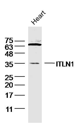
ITLN1 Polyclonal Antibody
Purified Rabbit Polyclonal Antibody (Pab)
- SPECIFICATION
- CITATIONS
- PROTOCOLS
- BACKGROUND

Application
| WB, IHC-P, IHC-F, IF, E |
|---|---|
| Primary Accession | Q8WWA0 |
| Reactivity | Rat, Pig, Bovine |
| Host | Rabbit |
| Clonality | Polyclonal |
| Calculated MW | 35 KDa |
| Physical State | Liquid |
| Immunogen | KLH conjugated synthetic peptide derived from human ITLN1 |
| Epitope Specificity | 131-230/313 |
| Isotype | IgG |
| Purity | affinity purified by Protein A |
| Buffer | 0.01M TBS (pH7.4) with 1% BSA, 0.02% Proclin300 and 50% Glycerol. |
| SUBCELLULAR LOCATION | Cell membrane; Lipid-anchor, GPI-anchor. Secreted. Note=Enriched in lipid rafts. |
| SIMILARITY | Contains 1 fibrinogen C-terminal domain. |
| SUBUNIT | Homotrimer; disulfide-linked. |
| Post-translational modifications | N-glycosylated. |
| Important Note | This product as supplied is intended for research use only, not for use in human, therapeutic or diagnostic applications. |
| Background Descriptions | ITLN1 (Intelectin 1) is a Protein Coding gene. Diseases associated with ITLN1 include Obesity and Diabetes Mellitus, Noninsulin-Dependent. Among its related pathways are Common Cytokine Receptor Gamma-Chain Family Signaling Pathways and Innate Immune System. GO annotations related to this gene include carbohydrate binding. An important paralog of this gene is ITLN2. |
| Gene ID | 55600 |
|---|---|
| Other Names | Intelectin-1, ITLN-1, Endothelial lectin HL-1, Galactofuranose-binding lectin, Intestinal lactoferrin receptor, Omentin, ITLN1, INTL, ITLN, LFR |
| Target/Specificity | Highly expressed in omental adipose tissue where it is found in stromal vascular cells but not in fat cells but is barely detectable in subcutaneous adipose tissue (at protein level). Highly expressed in the small intestine. Also found in the heart, testis, colon, salivary gland, skeletal muscle, pancreas and thyroid and, to a lesser degree, in the uterus, spleen, prostate, lymph node and thymus. |
| Dilution | WB=1:500-2000,IHC-P=1:100-500,IHC-F=1:100-500,IF=1:100-500,ELISA=1:5000-10000 |
| Storage | Store at -20 ℃ for one year. Avoid repeated freeze/thaw cycles. When reconstituted in sterile pH 7.4 0.01M PBS or diluent of antibody the antibody is stable for at least two weeks at 2-4 ℃. |
| Name | ITLN1 |
|---|---|
| Synonyms | INTL, ITLN, LFR |
| Function | Lectin that specifically recognizes microbial carbohydrate chains in a calcium-dependent manner (PubMed:11313366, PubMed:26148048). Binds to microbial glycans that contain a terminal acyclic 1,2-diol moiety, including beta-linked D-galactofuranose (beta- Galf), D-phosphoglycerol-modified glycans, D-glycero-D-talo-oct-2- ulosonic acid (KO) and 3-deoxy-D-manno-oct-2-ulosonic acid (KDO) (PubMed:26148048). Binds to glycans from Gram-positive and Gram- negative bacteria, including K.pneumoniae, S.pneumoniae, Y.pestis, P.mirabilis and P.vulgaris (PubMed:26148048). Does not bind human glycans (PubMed:26148048). Probably plays a role in the defense system against microorganisms (Probable). May function as adipokine that has no effect on basal glucose uptake but enhances insulin-stimulated glucose uptake in adipocytes (PubMed:16531507). Increases AKT phosphorylation in the absence and presence of insulin (PubMed:16531507). May interact with lactoferrin/LTF and increase its uptake, and may thereby play a role in iron absorption (PubMed:11747454, PubMed:23921499). |
| Cellular Location | Cell membrane; Lipid-anchor, GPI-anchor. Secreted. Note=Enriched in lipid rafts {ECO:0000250|UniProtKB:O88310} |
| Tissue Location | Highly expressed in omental adipose tissue where it is found in stromal vascular cells but not in fat cells but is barely detectable in subcutaneous adipose tissue (at protein level) (PubMed:16531507). Highly expressed in the small intestine. Also found in the heart, testis, colon, salivary gland, skeletal muscle, pancreas and thyroid and, to a lesser degree, in the uterus, spleen, prostate, lymph node and thymus. |
Research Areas
Citations (0)

Thousands of laboratories across the world have published research that depended on the performance of antibodies from Abcepta to advance their research. Check out links to articles that cite our products in major peer-reviewed journals, organized by research category.
Submit your citation using an Abcepta antibody to
info@abcepta.com, and receive a free "I Love Antibodies" mug.
info@abcepta.com, and receive a free "I Love Antibodies" mug.

Application Protocols
Provided below are standard protocols that you may find useful for product applications.
Abcepta welcomes feedback from its customers.
If you have used an Abcepta product and would like to share how it has performed, please click on the "Submit Review" button and provide the requested information. Our staff will examine and post your review and contact you if needed.
If you have any additional inquiries please email technical services at tech@abcepta.com.
$ 385.00
Cat# AP58286
Ordering Information
United States
AlbaniaAustraliaAustriaBelgiumBosnia & HerzegovinaBrazilBulgariaCanadaCentral AmericaChinaCroatiaCyprusCzech RepublicDenmarkEstoniaFinlandFranceGermanyGreeceHong KongHungaryIcelandIndiaIndonesiaIrelandIsraelItalyJapanLatviaLithuaniaLuxembourgMacedoniaMalaysiaMaltaMexicoNetherlandsNew ZealandNorwayPakistanPolandPortugalRomaniaSerbiaSingaporeSlovakiaSloveniaSouth AfricaSouth KoreaSpainSwedenSwitzerlandTaiwanTurkeyUnited KingdomUnited StatesVietnamWorldwideOthers
USA Headquarters
(888) 735-7227 / (858) 622-0099 or (858) 875-1900
Shipping Information
Domestic orders (in stock items)
Shipped out the same day. Orders placed after 1 PM (PST) will ship out the next business day.
International orders
Contact your local distributors
