


 Foundational characteristics of cancer include proliferation, angiogenesis, migration, evasion of apoptosis, and cellular immortality. Find key markers for these cellular processes and antibodies to detect them.
 The SUMOplot™ Analysis Program predicts and scores sumoylation sites in your protein. SUMOylation is a post-translational modification involved in various cellular processes, such as nuclear-cytosolic transport, transcriptional regulation, apoptosis, protein stability, response to stress, and progression through the cell cycle.
 The Autophagy Receptor Motif Plotter predicts and scores autophagy receptor binding sites in your protein. Identifying proteins connected to this pathway is critical to understanding the role of autophagy in physiological as well as pathological processes such as development, differentiation, neurodegenerative diseases, stress, infection, and cancer.
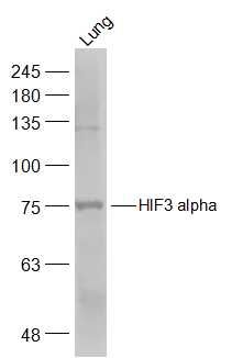
HIF3 alpha Polyclonal Antibody
Purified Rabbit Polyclonal Antibody (Pab)
- SPECIFICATION
- CITATIONS
- PROTOCOLS
- BACKGROUND

Application
| WB, IHC-P, IHC-F, IF, ICC, E |
|---|---|
| Primary Accession | Q9Y2N7 |
| Reactivity | Rat, Pig, Dog, Bovine |
| Host | Rabbit |
| Clonality | Polyclonal |
| Calculated MW | 74 KDa |
| Physical State | Liquid |
| Immunogen | KLH conjugated synthetic peptide derived from human HIF3 alpha |
| Epitope Specificity | 131-230/669 |
| Isotype | IgG |
| Purity | affinity purified by Protein A |
| Buffer | 0.01M TBS (pH7.4) with 1% BSA, 0.02% Proclin300 and 50% Glycerol. |
| SUBCELLULAR LOCATION | Nucleus. Cytoplasm. Note=In the nuclei of all periportal and perivenous hepatocytes. In the distal perivenous zone, detected in the cytoplasm of the hepatocytes. |
| SIMILARITY | Contains 1 basic helix-loop-helix (bHLH) domain.Contains 2 PAS (PER-ARNT-SIM) domains. |
| SUBUNIT | Heterodimerizes with ARNT. Interacts via the oxygen-dependent degradation domain (ODD) with the beta domain of VHL. |
| Important Note | This product as supplied is intended for research use only, not for use in human, therapeutic or diagnostic applications. |
| Background Descriptions | Hypoxia-inducible factor (HIF) is one of the most important factors in the cellular response to hypoxia, transcriptionally activating genes encoding proteins that mediate adaptive responses to reduced oxygen availability. HIF is a heterodimer consisting of one of three subunits, HIF1 alpha, HIF2 alpha, or HIF3 alpha. HIF target genes play critical roles in metabolism, angiogenesis, cell proliferation and cell survival. HIF3 alpha protein is one of several alpha/beta-subunit heterodimeric transcription factors that regulate many adaptive responses to low oxygen tension (hypoxia). The alpha 3 subunit lacks the transactivation domain found in factors containing either the alpha 1 or alpha 2 subunits. HIF3 alpha may be a marker for tumor growth and angiogenesis. |
| Gene ID | 64344 |
|---|---|
| Other Names | Hypoxia-inducible factor 3-alpha, HIF-3-alpha, HIF3-alpha, Basic-helix-loop-helix-PAS protein MOP7, Class E basic helix-loop-helix protein 17, bHLHe17, HIF3-alpha-1, Inhibitory PAS domain protein, IPAS, Member of PAS protein 7, PAS domain-containing protein 7, HIF3A (HGNC:15825), BHLHE17, MOP7, PASD7 |
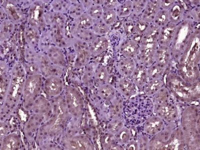
| Target/Specificity | Expressed in kidney. Expressed abundantly in lung epithelial cells. Expression is regulated in an oxygen-dependent manner. |
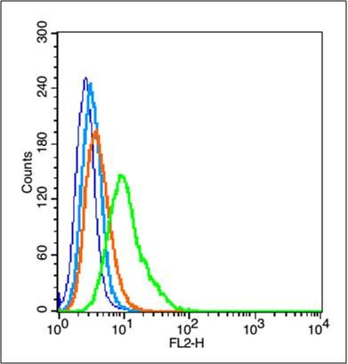
| Dilution | WB=1:500-2000,IHC-P=1:100-500,IHC-F=1:100-500,ICC=1:50,IF=1:100-500,Flow-Cyt=1 µg/Test,ELISA=1:5000-10000 |
| Format | 0.01M TBS(pH7.4), 0.09% (W/V) sodium azide and 50% Glyce |
| Storage | Store at -20 ℃ for one year. Avoid repeated freeze/thaw cycles. When reconstituted in sterile pH 7.4 0.01M PBS or diluent of antibody the antibody is stable for at least two weeks at 2-4 ℃. |
| Name | HIF3A (HGNC:15825) |
|---|---|
| Synonyms | BHLHE17, MOP7, PASD7 |
| Function | Acts as a transcriptional regulator in adaptive response to low oxygen tension. Acts as a regulator of hypoxia-inducible gene expression (PubMed:11573933, PubMed:16126907, PubMed:19694616, PubMed:20416395, PubMed:21069422). Functions as an inhibitor of angiogenesis in hypoxic cells of the cornea. Plays a role in the development of the cardiorespiratory system. May also be involved in apoptosis (By similarity). |
| Cellular Location | Nucleus. Cytoplasm Nucleus speckle {ECO:0000250|UniProtKB:Q0VBL6}. Mitochondrion {ECO:0000250|UniProtKB:Q0VBL6}. Note=In the nuclei of all periportal and perivenous hepatocytes. In the distal perivenous zone, detected in the cytoplasm of the hepatocytes. Shuttles between the nucleus and the cytoplasm in a CRM1-dependent manner. Colocalizes with BAD in the cytoplasm. Colocalizes with EPAS1 and HIF1A in the nucleus and speckles (By similarity). Localized in the cytoplasm and nuclei under normoxia, but increased in the nucleus under hypoxic conditions (PubMed:19694616). Colocalized with HIF1A in kidney tumors (PubMed:19694616). {ECO:0000250|UniProtKB:Q0VBL6, ECO:0000250|UniProtKB:Q9JHS2, ECO:0000269|PubMed:19694616} |
| Tissue Location | Expressed in vascular cells (at protein level) (PubMed:21069422). Expressed in kidney (PubMed:11573933, PubMed:19694616). Expressed in lung epithelial cells (PubMed:16775626) Expressed in endothelial cells (venous and arterial cells from umbilical cord and aortic endothelial cells) and in vascular smooth muscle cells (aorta) (PubMed:21069422). Strongly expressed in the heart, placenta, and skeletal muscle, whereas a weak expression profile was found in the lung, liver, and kidney (PubMed:12538644). Expressed weakly in cell renal cell carcinoma (CC-RCC) compared to normal renal cells (PubMed:16126907). Expression is down-regulated in numerous kidney tumor cells compared to non tumor kidney tissues (PubMed:16126907). Isoform 2 is expressed in heart, placenta, lung, liver, skeletal muscle and pancreas and in numerous cancer cell lines (PubMed:20416395). Isoform 3 and isoform 4 are weakly expressed in heart, placenta, lung, liver, skeletal muscle and pancreas (PubMed:20416395). Isoform 4 is expressed in fetal tissues, such as heart, brain, thymus, lung, liver, skeletal kidney and spleen (PubMed:20416395). Isoform 3 is weakly expressed in fetal tissues, such as liver and kidney (PubMed:20416395). |
Research Areas
Citations (0)

Thousands of laboratories across the world have published research that depended on the performance of antibodies from Abcepta to advance their research. Check out links to articles that cite our products in major peer-reviewed journals, organized by research category.
Submit your citation using an Abcepta antibody to
info@abcepta.com, and receive a free "I Love Antibodies" mug.
info@abcepta.com, and receive a free "I Love Antibodies" mug.

Application Protocols
Provided below are standard protocols that you may find useful for product applications.
Abcepta welcomes feedback from its customers.
If you have used an Abcepta product and would like to share how it has performed, please click on the "Submit Review" button and provide the requested information. Our staff will examine and post your review and contact you if needed.
If you have any additional inquiries please email technical services at tech@abcepta.com.
$ 385.00
Cat# AP58343
Ordering Information
United States
AlbaniaAustraliaAustriaBelgiumBosnia & HerzegovinaBrazilBulgariaCanadaCentral AmericaChinaCroatiaCyprusCzech RepublicDenmarkEstoniaFinlandFranceGermanyGreeceHong KongHungaryIcelandIndiaIndonesiaIrelandIsraelItalyJapanLatviaLithuaniaLuxembourgMacedoniaMalaysiaMaltaMexicoNetherlandsNew ZealandNorwayPakistanPolandPortugalRomaniaSerbiaSingaporeSlovakiaSloveniaSouth AfricaSouth KoreaSpainSwedenSwitzerlandTaiwanTurkeyUnited KingdomUnited StatesVietnamWorldwideOthers
USA Headquarters
(888) 735-7227 / (858) 622-0099 or (858) 875-1900
Other Products
Shipping Information
Domestic orders (in stock items)
Shipped out the same day. Orders placed after 1 PM (PST) will ship out the next business day.
International orders
Contact your local distributors
