


 Foundational characteristics of cancer include proliferation, angiogenesis, migration, evasion of apoptosis, and cellular immortality. Find key markers for these cellular processes and antibodies to detect them.
 The SUMOplot™ Analysis Program predicts and scores sumoylation sites in your protein. SUMOylation is a post-translational modification involved in various cellular processes, such as nuclear-cytosolic transport, transcriptional regulation, apoptosis, protein stability, response to stress, and progression through the cell cycle.
 The Autophagy Receptor Motif Plotter predicts and scores autophagy receptor binding sites in your protein. Identifying proteins connected to this pathway is critical to understanding the role of autophagy in physiological as well as pathological processes such as development, differentiation, neurodegenerative diseases, stress, infection, and cancer.
CRISP3 Polyclonal Antibody
Purified Rabbit Polyclonal Antibody (Pab)
- SPECIFICATION
- CITATIONS
- PROTOCOLS
- BACKGROUND

Application
| WB, IHC-P, IHC-F, IF, E |
|---|---|
| Primary Accession | P54108 |
| Reactivity | Rat, Dog, Bovine |
| Host | Rabbit |
| Clonality | Polyclonal |
| Calculated MW | 25 KDa |
| Physical State | Liquid |
| Immunogen | KLH conjugated synthetic peptide derived from human CRISP3 |
| Epitope Specificity | 21-120/245 |
| Isotype | IgG |
| Purity | affinity purified by Protein A |
| Buffer | 0.01M TBS (pH7.4) with 1% BSA, 0.02% Proclin300 and 50% Glycerol. |
| SUBCELLULAR LOCATION | Secreted. In neutrophils, localized in specific granules. |
| SIMILARITY | Belongs to the CRISP family. |
| SUBUNIT | Interacts with A1BG. |
| Important Note | This product as supplied is intended for research use only, not for use in human, therapeutic or diagnostic applications. |
| Background Descriptions | Cysteine-rich secretory proteins (CRISPs) represent a family of evolutionarily conserved proteins which may play a role in the innate immune system and are transcriptionally regulated by androgens in several tissues. AEG is a sperm surface protein involved in the fusion of egg and sperm. Although CRISP-1 (also designated AEG-like protein, ARP, cysteine-rich secretory protein-1 or AEG-related protein) is not the ortholog of rodent AEG, it resembles AEG in that it is an epididymal secretory glycoprotein that binds to the postacrosomal region of the sperm head. CRISP-1 coats the postacrosomal region of sperm heads as they pass through the epididymis. CRISP-1 is found in all regions of the epididymis, ductus deferens, seminal plasma and sperm. CRISP-3 is expressed in pancreas and prostate tissues and, along with CRISP-1, is expressed in saliva. The gene that encodes CRISP-3 is an early response gene that may participate in the pathophysiology of the autoimmune lesions of Sjogren’s syndrome. |
| Gene ID | 10321 |
|---|---|
| Other Names | Cysteine-rich secretory protein 3, CRISP-3, Specific granule protein of 28 kDa, SGP28, CRISP3 |
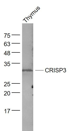
| Target/Specificity | Salivary gland, pancreas and prostate epididymis, ovary, thymus and colon. |
| Dilution | WB=1:500-2000,IHC-P=1:100-500,IHC-F=1:100-500,IF=1:100-500,ELISA=1:5000-10000 |
| Storage | Store at -20 ℃ for one year. Avoid repeated freeze/thaw cycles. When reconstituted in sterile pH 7.4 0.01M PBS or diluent of antibody the antibody is stable for at least two weeks at 2-4 ℃. |
| Name | CRISP3 |
|---|---|
| Cellular Location | Secreted. Note=In neutrophils, localized in specific granules |
| Tissue Location | Salivary gland, pancreas and prostate > epididymis, ovary, thymus and colon |
Research Areas
Citations (0)

Thousands of laboratories across the world have published research that depended on the performance of antibodies from Abcepta to advance their research. Check out links to articles that cite our products in major peer-reviewed journals, organized by research category.
Submit your citation using an Abcepta antibody to
info@abcepta.com, and receive a free "I Love Antibodies" mug.
info@abcepta.com, and receive a free "I Love Antibodies" mug.

Application Protocols
Provided below are standard protocols that you may find useful for product applications.
Abcepta welcomes feedback from its customers.
If you have used an Abcepta product and would like to share how it has performed, please click on the "Submit Review" button and provide the requested information. Our staff will examine and post your review and contact you if needed.
If you have any additional inquiries please email technical services at tech@abcepta.com.
$ 385.00
Cat# AP58459
Ordering Information
United States
AlbaniaAustraliaAustriaBelgiumBosnia & HerzegovinaBrazilBulgariaCanadaCentral AmericaChinaCroatiaCyprusCzech RepublicDenmarkEstoniaFinlandFranceGermanyGreeceHong KongHungaryIcelandIndiaIndonesiaIrelandIsraelItalyJapanLatviaLithuaniaLuxembourgMacedoniaMalaysiaMaltaMexicoNetherlandsNew ZealandNorwayPakistanPolandPortugalRomaniaSerbiaSingaporeSlovakiaSloveniaSouth AfricaSouth KoreaSpainSwedenSwitzerlandTaiwanTurkeyUnited KingdomUnited StatesVietnamWorldwideOthers
USA Headquarters
(888) 735-7227 / (858) 622-0099 or (858) 875-1900
Other Products
Shipping Information
Domestic orders (in stock items)
Shipped out the same day. Orders placed after 1 PM (PST) will ship out the next business day.
International orders
Contact your local distributors
