


 Foundational characteristics of cancer include proliferation, angiogenesis, migration, evasion of apoptosis, and cellular immortality. Find key markers for these cellular processes and antibodies to detect them.
 The SUMOplot™ Analysis Program predicts and scores sumoylation sites in your protein. SUMOylation is a post-translational modification involved in various cellular processes, such as nuclear-cytosolic transport, transcriptional regulation, apoptosis, protein stability, response to stress, and progression through the cell cycle.
 The Autophagy Receptor Motif Plotter predicts and scores autophagy receptor binding sites in your protein. Identifying proteins connected to this pathway is critical to understanding the role of autophagy in physiological as well as pathological processes such as development, differentiation, neurodegenerative diseases, stress, infection, and cancer.
HSD17B6 Polyclonal Antibody
Purified Rabbit Polyclonal Antibody (Pab)
- SPECIFICATION
- CITATIONS
- PROTOCOLS
- BACKGROUND

Application
| WB, IHC-P, IHC-F, IF, E |
|---|---|
| Primary Accession | O14756 |
| Reactivity | Rat, Pig |
| Host | Rabbit |
| Clonality | Polyclonal |
| Calculated MW | 33 KDa |
| Physical State | Liquid |
| Immunogen | KLH conjugated synthetic peptide derived from human HSD17B6 |
| Epitope Specificity | 61-160/317 |
| Isotype | IgG |
| Purity | affinity purified by Protein A |
| Buffer | 0.01M TBS (pH7.4) with 1% BSA, 0.02% Proclin300 and 50% Glycerol. |
| SUBCELLULAR LOCATION | Microsome membrane; Peripheral membrane protein; Lumenal side. Early endosome membrane; Peripheral membrane protein; Lumenal side (Potential). |
| SIMILARITY | Belongs to the short-chain dehydrogenases/reductases (SDR) family. |
| Important Note | This product as supplied is intended for research use only, not for use in human, therapeutic or diagnostic applications. |
| Background Descriptions | The protein encoded by this gene has both oxidoreductase and epimerase activities and is involved in androgen catabolism. The oxidoreductase activity can convert 3 alpha-adiol to dihydrotestosterone, while the epimerase activity can convert androsterone to epi-androsterone. Both reactions use NAD+ as the preferred cofactor. This gene is a member of the retinol dehydrogenase family. [provided by RefSeq, Aug 2013] |
| Gene ID | 8630 |
|---|---|
| Other Names | 17-beta-hydroxysteroid dehydrogenase type 6, 17-beta-HSD 6, 17-beta-HSD6, 1.1.1.105, 1.1.1.209, 1.1.1.239, 3-alpha->beta-hydroxysteroid epimerase, 3-alpha->beta-HSE, Oxidative 3-alpha hydroxysteroid dehydrogenase, Short chain dehydrogenase/reductase family 9C member 6, HSD17B6, RODH, SDR9C6 |
| Target/Specificity | Detected in liver and prostate (at protein level). Detected in adult liver, lung, brain, placenta, prostate, adrenal gland, testis, mammary gland, spleen, spinal cord and uterus. Detected in caudate nucleus, and at lower levels in amygdala, corpus callosum, hippocampus, substantia nigra and thalamus. Detected in fetal lung, liver and brain. |
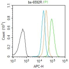
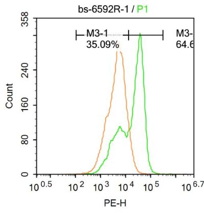
| Dilution | WB=1:500-2000,IHC-P=1:100-500,IHC-F=1:100-500,IF=1:100-500,Flow-Cyt=1ug/Test,ELISA=1:5000-10000 |
| Storage | Store at -20 ℃ for one year. Avoid repeated freeze/thaw cycles. When reconstituted in sterile pH 7.4 0.01M PBS or diluent of antibody the antibody is stable for at least two weeks at 2-4 ℃. |
| Name | HSD17B6 |
|---|---|
| Synonyms | RODH, SDR9C6 |
| Function | NAD-dependent oxidoreductase with broad substrate specificity that shows both oxidative and reductive activity (in vitro). Has 17- beta-hydroxysteroid dehydrogenase activity towards various steroids (in vitro). Converts 5-alpha-androstan-3-alpha,17-beta-diol to androsterone and estradiol to estrone (in vitro). Has 3-alpha-hydroxysteroid dehydrogenase activity towards androsterone (in vitro). Has retinol dehydrogenase activity towards all-trans-retinol (in vitro). Can convert androsterone to epi-androsterone. Androsterone is first oxidized to 5-alpha-androstane-3,17-dione and then reduced to epi- andosterone. Can act on both C-19 and C-21 3-alpha-hydroxysteroids. |
| Cellular Location | Microsome membrane; Peripheral membrane protein; Lumenal side. Early endosome membrane; Peripheral membrane protein; Lumenal side |
| Tissue Location | Detected in liver and prostate (at protein level). Detected in adult liver, lung, brain, placenta, prostate, adrenal gland, testis, mammary gland, spleen, spinal cord and uterus. Detected in caudate nucleus, and at lower levels in amygdala, corpus callosum, hippocampus, substantia nigra and thalamus. Detected in fetal lung, liver and brain. |
Research Areas
Citations (0)

Thousands of laboratories across the world have published research that depended on the performance of antibodies from Abcepta to advance their research. Check out links to articles that cite our products in major peer-reviewed journals, organized by research category.
Submit your citation using an Abcepta antibody to
info@abcepta.com, and receive a free "I Love Antibodies" mug.
info@abcepta.com, and receive a free "I Love Antibodies" mug.

Application Protocols
Provided below are standard protocols that you may find useful for product applications.
Abcepta welcomes feedback from its customers.
If you have used an Abcepta product and would like to share how it has performed, please click on the "Submit Review" button and provide the requested information. Our staff will examine and post your review and contact you if needed.
If you have any additional inquiries please email technical services at tech@abcepta.com.
$ 385.00
Cat# AP58477
Ordering Information
United States
AlbaniaAustraliaAustriaBelgiumBosnia & HerzegovinaBrazilBulgariaCanadaCentral AmericaChinaCroatiaCyprusCzech RepublicDenmarkEstoniaFinlandFranceGermanyGreeceHong KongHungaryIcelandIndiaIndonesiaIrelandIsraelItalyJapanLatviaLithuaniaLuxembourgMacedoniaMalaysiaMaltaMexicoNetherlandsNew ZealandNorwayPakistanPolandPortugalRomaniaSerbiaSingaporeSlovakiaSloveniaSouth AfricaSouth KoreaSpainSwedenSwitzerlandTaiwanTurkeyUnited KingdomUnited StatesVietnamWorldwideOthers
USA Headquarters
(888) 735-7227 / (858) 622-0099 or (858) 875-1900
Shipping Information
Domestic orders (in stock items)
Shipped out the same day. Orders placed after 1 PM (PST) will ship out the next business day.
International orders
Contact your local distributors
