


 Foundational characteristics of cancer include proliferation, angiogenesis, migration, evasion of apoptosis, and cellular immortality. Find key markers for these cellular processes and antibodies to detect them.
 The SUMOplot™ Analysis Program predicts and scores sumoylation sites in your protein. SUMOylation is a post-translational modification involved in various cellular processes, such as nuclear-cytosolic transport, transcriptional regulation, apoptosis, protein stability, response to stress, and progression through the cell cycle.
 The Autophagy Receptor Motif Plotter predicts and scores autophagy receptor binding sites in your protein. Identifying proteins connected to this pathway is critical to understanding the role of autophagy in physiological as well as pathological processes such as development, differentiation, neurodegenerative diseases, stress, infection, and cancer.
Netrin G1 ligand Polyclonal Antibody
Purified Rabbit Polyclonal Antibody (Pab)
- SPECIFICATION
- CITATIONS
- PROTOCOLS
- BACKGROUND

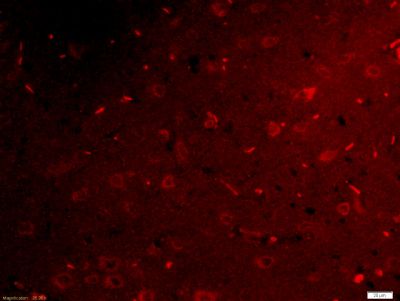
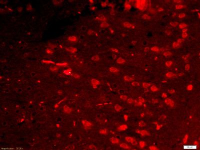
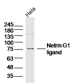
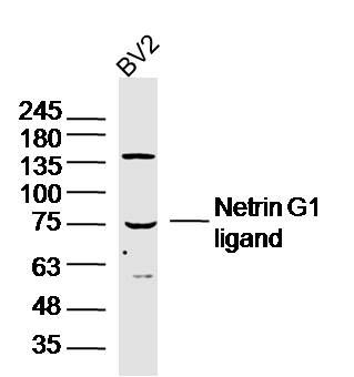
Application
| WB, IHC-P, IHC-F, IF, E |
|---|---|
| Primary Accession | Q9HCJ2 |
| Reactivity | Rat, Dog, Bovine |
| Host | Rabbit |
| Clonality | Polyclonal |
| Calculated MW | 70 KDa |
| Physical State | Liquid |
| Immunogen | KLH conjugated synthetic peptide derived from human Netrin G1 ligand |
| Epitope Specificity | 561-640/640 |
| Isotype | IgG |
| Purity | affinity purified by Protein A |
| Buffer | 0.01M TBS (pH7.4) with 1% BSA, 0.02% Proclin300 and 50% Glycerol. |
| SUBCELLULAR LOCATION | Membrane. |
| SIMILARITY | Contains 1 Ig-like C2-type (immunoglobulin-like) domain.Contains 9 LRR (leucine-rich) repeats.Contains 1 LRRCT domain.Contains 1 LRRNT domain. |
| SUBUNIT | Interacts with NTNG1 and WHRN. |
| Important Note | This product as supplied is intended for research use only, not for use in human, therapeutic or diagnostic applications. |
| Background Descriptions | NGL-1 is a single pass type I membrane protein that acts as a cell adhesion molecule. It contains nine leucine-rich repeats (LRR) and one Ig-like C2-type domain. NGL-1 is predominantly expressed in the striatum and the cerebral cortex of both the embryonic and adult brain. NGL-1 specifically interacts with Netrin G1 (a molecule involved in axon guidance in the developing central nervous system) via its LRR region. NGL-1 plays a role in the regulation of neurite outgrowth of developing thalamic neurons. Soluble NGL-1 inhibits thalamic axon outgrowth while NGL-1 that is bound to the surface of developing thalamocortical axons stimulates growth. NGL-1 also interacts with Whirlin possibly stablizing interstereociliar links. |
| Gene ID | 57689 |
|---|---|
| Other Names | Leucine-rich repeat-containing protein 4C, Netrin-G1 ligand, NGL-1, LRRC4C, KIAA1580, NGL1 |
| Target/Specificity | Highly expressed in the cerebral cortex, including frontal, parietal and occipital lobes. Putamen, amygdala, hippocampus and medulla oblongata show moderate expression. Caudate nucleus and thalamus express small amounts, whereas other brain regions show very weak or no expression. |
| Dilution | WB=1:500-2000,IHC-P=1:100-500,IHC-F=1:100-500,IF=1:100-500,ELISA=1:5000-10000 |
| Storage | Store at -20 ℃ for one year. Avoid repeated freeze/thaw cycles. When reconstituted in sterile pH 7.4 0.01M PBS or diluent of antibody the antibody is stable for at least two weeks at 2-4 ℃. |
| Name | LRRC4C |
|---|---|
| Synonyms | KIAA1580, NGL1 |
| Function | May promote neurite outgrowth of developing thalamic neurons. |
| Cellular Location | Postsynaptic cell membrane; Single-pass type I membrane protein |
| Tissue Location | Highly expressed in the cerebral cortex, including frontal, parietal and occipital lobes. Putamen, amygdala, hippocampus and medulla oblongata show moderate expression. Caudate nucleus and thalamus express small amounts, whereas other brain regions show very weak or no expression. |
Research Areas
Citations (0)

Thousands of laboratories across the world have published research that depended on the performance of antibodies from Abcepta to advance their research. Check out links to articles that cite our products in major peer-reviewed journals, organized by research category.
Submit your citation using an Abcepta antibody to
info@abcepta.com, and receive a free "I Love Antibodies" mug.
info@abcepta.com, and receive a free "I Love Antibodies" mug.

Application Protocols
Provided below are standard protocols that you may find useful for product applications.
Abcepta welcomes feedback from its customers.
If you have used an Abcepta product and would like to share how it has performed, please click on the "Submit Review" button and provide the requested information. Our staff will examine and post your review and contact you if needed.
If you have any additional inquiries please email technical services at tech@abcepta.com.
$ 385.00
Cat# AP58495
Ordering Information
United States
AlbaniaAustraliaAustriaBelgiumBosnia & HerzegovinaBrazilBulgariaCanadaCentral AmericaChinaCroatiaCyprusCzech RepublicDenmarkEstoniaFinlandFranceGermanyGreeceHong KongHungaryIcelandIndiaIndonesiaIrelandIsraelItalyJapanLatviaLithuaniaLuxembourgMacedoniaMalaysiaMaltaMexicoNetherlandsNew ZealandNorwayPakistanPolandPortugalRomaniaSerbiaSingaporeSlovakiaSloveniaSouth AfricaSouth KoreaSpainSwedenSwitzerlandTaiwanTurkeyUnited KingdomUnited StatesVietnamWorldwideOthers
USA Headquarters
(888) 735-7227 / (858) 622-0099 or (858) 875-1900
Shipping Information
Domestic orders (in stock items)
Shipped out the same day. Orders placed after 1 PM (PST) will ship out the next business day.
International orders
Contact your local distributors
