


 Foundational characteristics of cancer include proliferation, angiogenesis, migration, evasion of apoptosis, and cellular immortality. Find key markers for these cellular processes and antibodies to detect them.
 The SUMOplot™ Analysis Program predicts and scores sumoylation sites in your protein. SUMOylation is a post-translational modification involved in various cellular processes, such as nuclear-cytosolic transport, transcriptional regulation, apoptosis, protein stability, response to stress, and progression through the cell cycle.
 The Autophagy Receptor Motif Plotter predicts and scores autophagy receptor binding sites in your protein. Identifying proteins connected to this pathway is critical to understanding the role of autophagy in physiological as well as pathological processes such as development, differentiation, neurodegenerative diseases, stress, infection, and cancer.
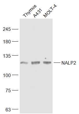
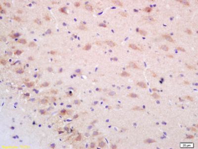
NALP2 Polyclonal Antibody
Purified Rabbit Polyclonal Antibody (Pab)
- SPECIFICATION
- CITATIONS
- PROTOCOLS
- BACKGROUND

Application
| WB, IHC-P, IHC-F, IF, ICC, E |
|---|---|
| Primary Accession | Q9NX02 |
| Reactivity | Rat |
| Host | Rabbit |
| Clonality | Polyclonal |
| Calculated MW | 117 KDa |
| Physical State | Liquid |
| Immunogen | KLH conjugated synthetic peptide derived from human NACHT, LRR and PYD domains-containing protein 2 |
| Epitope Specificity | 851-1062/1062 |
| Isotype | IgG |
| Purity | affinity purified by Protein A |
| Buffer | 0.01M TBS (pH7.4) with 1% BSA, 0.02% Proclin300 and 50% Glycerol. |
| SUBCELLULAR LOCATION | Cytoplasm. |
| SIMILARITY | Belongs to the NLRP family.Contains 1 DAPIN domain. Contains 8 LRR (leucine-rich) repeats. Contains 1 NACHT domain. |
| SUBUNIT | Interacts with CHUK, IKBKB and IKBKG, as well as with full-length PYCARD and with the DAPIN domain of NAPL1, but not the full-length protein. |
| Important Note | This product as supplied is intended for research use only, not for use in human, therapeutic or diagnostic applications. |
| Background Descriptions | NALP proteins, such as NALP2, are characterized by an N-terminal pyrin (MIM 608107) domain (PYD) and are involved in the activation of caspase-1 (CASP1; MIM 147678) by Toll-like receptors (see TLR4; MIM 603030). They may also be involved in protein complexes that activate proinflammatory caspases (Tschopp et al., 2003 [PubMed 12563287]).[supplied by OMIM, Mar 2008]. |
| Gene ID | 55655 |
|---|---|
| Other Names | NACHT, LRR and PYD domains-containing protein 2, Nucleotide-binding site protein 1, PYRIN domain and NACHT domain-containing protein 1, PYRIN-containing APAF1-like protein 2, NLRP2, NALP2, NBS1, PAN1, PYPAF2 |
| Target/Specificity | Expressed at high levels in lung, placenta and thymus and at lower levels in ovary, intestine and brain. |
| Dilution | WB=1:500-2000,IHC-P=1:100-500,IHC-F=1:100-500,ICC=1:100-500,IF=1:100-500,ELISA=1:5000-10000 |
| Storage | Store at -20 ℃ for one year. Avoid repeated freeze/thaw cycles. When reconstituted in sterile pH 7.4 0.01M PBS or diluent of antibody the antibody is stable for at least two weeks at 2-4 ℃. |
| Name | NLRP2 |
|---|---|
| Synonyms | NALP2, NBS1, PAN1, PYPAF2 |
| Function | Suppresses TNF- and CD40-induced NFKB1 activity at the level of the IKK complex, by inhibiting NFKBIA degradation induced by TNF. When associated with PYCARD, activates CASP1, leading to the secretion of mature pro-inflammatory cytokine IL1B. May be a component of the inflammasome, a protein complex which also includes PYCARD, CARD8 and CASP1 and whose function would be the activation of pro-inflammatory caspases. |
| Cellular Location | Cytoplasm |
| Tissue Location | Expressed at high levels in lung, placenta and thymus and at lower levels in ovary, intestine and brain (PubMed:15456791). Highly abundant in oocytes and early embryos, however poorly expressed in somatic tissues such as brain, kidney, liver and spinal cord (PubMed:30877238). |
Research Areas
Citations (0)

Thousands of laboratories across the world have published research that depended on the performance of antibodies from Abcepta to advance their research. Check out links to articles that cite our products in major peer-reviewed journals, organized by research category.
Submit your citation using an Abcepta antibody to
info@abcepta.com, and receive a free "I Love Antibodies" mug.
info@abcepta.com, and receive a free "I Love Antibodies" mug.

Application Protocols
Provided below are standard protocols that you may find useful for product applications.
Abcepta welcomes feedback from its customers.
If you have used an Abcepta product and would like to share how it has performed, please click on the "Submit Review" button and provide the requested information. Our staff will examine and post your review and contact you if needed.
If you have any additional inquiries please email technical services at tech@abcepta.com.
$ 385.00
Cat# AP58498
Ordering Information
United States
AlbaniaAustraliaAustriaBelgiumBosnia & HerzegovinaBrazilBulgariaCanadaCentral AmericaChinaCroatiaCyprusCzech RepublicDenmarkEstoniaFinlandFranceGermanyGreeceHong KongHungaryIcelandIndiaIndonesiaIrelandIsraelItalyJapanLatviaLithuaniaLuxembourgMacedoniaMalaysiaMaltaMexicoNetherlandsNew ZealandNorwayPakistanPolandPortugalRomaniaSerbiaSingaporeSlovakiaSloveniaSouth AfricaSouth KoreaSpainSwedenSwitzerlandTaiwanTurkeyUnited KingdomUnited StatesVietnamWorldwideOthers
USA Headquarters
(888) 735-7227 / (858) 622-0099 or (858) 875-1900
Other Products
Shipping Information
Domestic orders (in stock items)
Shipped out the same day. Orders placed after 1 PM (PST) will ship out the next business day.
International orders
Contact your local distributors
