


 Foundational characteristics of cancer include proliferation, angiogenesis, migration, evasion of apoptosis, and cellular immortality. Find key markers for these cellular processes and antibodies to detect them.
 The SUMOplot™ Analysis Program predicts and scores sumoylation sites in your protein. SUMOylation is a post-translational modification involved in various cellular processes, such as nuclear-cytosolic transport, transcriptional regulation, apoptosis, protein stability, response to stress, and progression through the cell cycle.
 The Autophagy Receptor Motif Plotter predicts and scores autophagy receptor binding sites in your protein. Identifying proteins connected to this pathway is critical to understanding the role of autophagy in physiological as well as pathological processes such as development, differentiation, neurodegenerative diseases, stress, infection, and cancer.
CARD7/NALP1 Polyclonal Antibody
Purified Rabbit Polyclonal Antibody (Pab)
- SPECIFICATION
- CITATIONS
- PROTOCOLS
- BACKGROUND

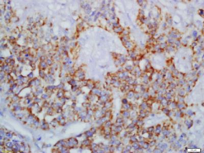
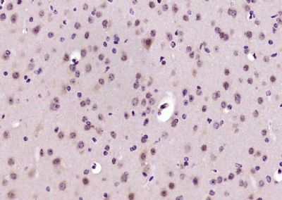
Application
| IHC-P, IHC-F, IF, E |
|---|---|
| Primary Accession | Q9C000 |
| Reactivity | Rat |
| Host | Rabbit |
| Clonality | Polyclonal |
| Calculated MW | 162 KDa |
| Physical State | Liquid |
| Immunogen | KLH conjugated synthetic peptide derived from human CARD7/NALP1 |
| Epitope Specificity | 301-400/1473 |
| Isotype | IgG |
| Purity | affinity purified by Protein A |
| Buffer | 0.01M TBS (pH7.4) with 1% BSA, 0.02% Proclin300 and 50% Glycerol. |
| SUBCELLULAR LOCATION | Cytoplasm. Nucleus. |
| SIMILARITY | Belongs to the NLRP family. Contains 1 CARD domain. Contains 1 DAPIN domain. Contains 6 LRR (leucine-rich) repeats. Contains 1 NACHT domain. |
| SUBUNIT | Interacts strongly with caspase-2, weakly with caspase-9 and with APAF1 in a cytochrome c-inducible way, leading to the formation of an apoptosome. This interaction may be ATP-dependent. Part of the NALP1 inflammasome complex which is involved in activation of caspase-1 and caspase-5, leading to processing of IL1B and IL18. The complex is activated by bacterial muramyl dipeptide which triggers ATP-binding and oligomerization of NALP1. |
| DISEASE | Genetic variations in NLRP1 are associated with susceptibility to vitiligo (VTLG) [MIM:193200]. VTLG is a pigmentary disorder of the skin characterized by circumscribed depigmented macules and patches, commonly on extensor aspects of extremities, on the face or neck and in skin folds. It is a progressive disorder in which some or all of the melanocytes in the affected skin are selectively destroyed. It is a multifactorial disorder with a complex etiology probably including autoimmune mechanisms, and is associated with an elevated risk of other autoimmune diseases.Genetic variations in NLRP1 gene are associated with susceptibility to vitiligo-associated multiple autoimmune disease type 1 (VAMAS1) [MIM:606579]. VAMAS1 is an autoimmune disorder characterized by the association of vitiligo with several autoimmune and autoinflammatory diseases including autoimmune thyroid disease, rheumatoid arthritis and systemic lupus erythematosus. |
| Important Note | This product as supplied is intended for research use only, not for use in human, therapeutic or diagnostic applications. |
| Background Descriptions | This gene encodes a member of the Ced-4 family of apoptosis proteins. Ced-family members contain a caspase recruitment domain (CARD) and are known to be key mediators of programmed cell death. The encoded protein contains a distinct N-terminal pyrin-like motif, which is possibly involved in protein-protein interactions. This protein interacts strongly with caspase 2 and weakly with caspase 9. Overexpression of this gene was demonstrated to induce apoptosis in cells. Multiple alternatively spliced transcript variants encoding distinct isoforms have been found for this gene, but the biological validity of some variants has not been determined. [provided by RefSeq, Jul 2008]. |
| Gene ID | 22861 |
|---|---|
| Other Names | NACHT, LRR and PYD domains-containing protein 1, 3.6.4.-, Caspase recruitment domain-containing protein 7, Death effector filament-forming ced-4-like apoptosis protein, Nucleotide-binding domain and caspase recruitment domain, NLRP1 (HGNC:14374) |
| Target/Specificity | Widely expressed. Isoform 1 and isoform 2 are expressed in peripheral blood leukocytes and chronic myelogenous leukemia cell line K-562, followed by thymus, spleen and heart. Also detected in brain, lung, placenta, small intestine, colon, kidney, liver, muscle, testis and epithelial cells. Absent from hematopoietic progenitor cells but expressed upon differentiation of cells into granulocytes and, to a lesser extent, monocytes. In peripheral blood cells, highest levels are found in T-lymphocytes, granulocytes and monocytes. Expression is significantly increased in bone marrow blast cells of some acute leukemia patients but not in solid tumors. |
| Dilution | IHC-P=1:100-500,IHC-F=1:100-500,IF=1:100-500,ELISA=1:5000-10000 |
| Format | 0.01M TBS(pH7.4), 0.09% (W/V) sodium azide and 50% Glyce |
| Storage | Store at -20 ℃ for one year. Avoid repeated freeze/thaw cycles. When reconstituted in sterile pH 7.4 0.01M PBS or diluent of antibody the antibody is stable for at least two weeks at 2-4 ℃. |
| Name | NLRP1 {ECO:0000303|PubMed:22665479, ECO:0000312|HGNC:HGNC:14374} |
|---|---|
| Function | Acts as the sensor component of the NLRP1 inflammasome, which mediates inflammasome activation in response to various pathogen- associated signals, leading to subsequent pyroptosis (PubMed:12191486, PubMed:17349957, PubMed:22665479, PubMed:27662089, PubMed:31484767, PubMed:33093214, PubMed:33410748, PubMed:33731929, PubMed:33731932, PubMed:35857590). Inflammasomes are supramolecular complexes that assemble in the cytosol in response to pathogens and other damage- associated signals and play critical roles in innate immunity and inflammation (PubMed:12191486, PubMed:17349957, PubMed:22665479). Acts as a recognition receptor (PRR): recognizes specific pathogens and other damage-associated signals, such as cleavage by some human enteroviruses and rhinoviruses, double-stranded RNA, UV-B irradiation, or Val-boroPro inhibitor, and mediates the formation of the inflammasome polymeric complex composed of NLRP1, CASP1 and PYCARD/ASC (PubMed:12191486, PubMed:17349957, PubMed:22665479, PubMed:25562666, PubMed:30096351, PubMed:30291141, PubMed:33093214, PubMed:33243852, PubMed:33410748, PubMed:35857590). In response to pathogen-associated signals, the N-terminal part of NLRP1 is degraded by the proteasome, releasing the cleaved C-terminal part of the protein (NACHT, LRR and PYD domains-containing protein 1, C-terminus), which polymerizes and associates with PYCARD/ASC to initiate the formation of the inflammasome complex: the NLRP1 inflammasome recruits pro-caspase-1 (proCASP1) and promotes caspase-1 (CASP1) activation, which subsequently cleaves and activates inflammatory cytokines IL1B and IL18 and gasdermin-D (GSDMD), leading to pyroptosis (PubMed:12191486, PubMed:17349957, PubMed:22665479, PubMed:32051255, PubMed:33093214). In the absence of GSDMD expression, the NLRP1 inflammasome is able to recruit and activate CASP8, leading to activation of gasdermin-E (GSDME) (PubMed:33852854, PubMed:35594856). Activation of NLRP1 inflammasome is also required for HMGB1 secretion; the active cytokines and HMGB1 stimulate inflammatory responses (PubMed:22801494). Binds ATP and shows ATPase activity (PubMed:11113115, PubMed:15212762, PubMed:33243852). Plays an important role in antiviral immunity and inflammation in the human airway epithelium (PubMed:33093214). Specifically recognizes a number of pathogen-associated signals: upon infection by human rhinoviruses 14 and 16 (HRV-14 and HRV-16), NLRP1 is cleaved and activated which triggers NLRP1-dependent inflammasome activation and IL18 secretion (PubMed:33093214). Positive-strand RNA viruses, such as Semliki forest virus and long dsRNA activate the NLRP1 inflammasome, triggering IL1B release in a NLRP1-dependent fashion (PubMed:33243852). Acts as a direct sensor for long dsRNA and thus RNA virus infection (PubMed:33243852). May also be activated by muramyl dipeptide (MDP), a fragment of bacterial peptidoglycan, in a NOD2- dependent manner (PubMed:18511561). The NLRP1 inflammasome is also activated in response to UV-B irradiation causing ribosome collisions: ribosome collisions cause phosphorylation and activation of NLRP1 in a MAP3K20-dependent manner, leading to pyroptosis (PubMed:35857590). |
| Cellular Location | Cytoplasm, cytosol. Cytoplasm. Nucleus. Note=Nucleocytoplasmic distribution in lymphoid organs (probably in T-cells) and in neurons. In epithelial cells, predominantly cytoplasmic. [NACHT, LRR and PYD domains-containing protein 1, N-terminus]: Nucleus. Note=(Microbial infection) Interaction with human herpes virus 8/HHV-8 proteins ORF45 promotes translocation of the N-terminal part of NLRP1 into the nucleus, relieving autoinhibition of the NLRP1 inflammasome and leading to its activation. |
| Tissue Location | Widely expressed (PubMed:11113115, PubMed:17164409). Abundantly expressed in primary immune cells (isoform 1 and isoform 2), including in neutrophils, monocytes/macrophages, dendritic cells (mostly Langerhans cells), and B- and T-lymphocytes (at protein level) (PubMed:15285719, PubMed:17164409). Strongly expressed in epithelial cells lining the glandular epithelium, such as that of the gastrointestinal tract (stomach, small intestine, colon), the respiratory tract (trachea and bronchi), and the endometrial and endocervical glands, gallbladder, prostate, and breast (at protein level). In testis, expressed in spermatogonia and primary spermatocytes, but not in Sertoli cells (at protein level). In the brain, expressed in neurons, in particular in pyramidal ones and in oligodendrocytes, but not detected in microglia (at protein level) (PubMed:17164409). Expressed in adult and fetal ocular tissues, including in adult and 24-week old fetal choroid, sclera, cornea, and optic nerve, as well as in adult retina and fetal retina/retinal pigment epithelium (PubMed:23349227). Highly expressed in the skin throughout the epidermis and in dermal fibroblasts, in both glabrous skin and plantar skin. It is detected in keratinocytes, but not in melanocytes. Expressed in epidermal appendages such as hair follicles (PubMed:27662089). |
Research Areas
Citations (0)

Thousands of laboratories across the world have published research that depended on the performance of antibodies from Abcepta to advance their research. Check out links to articles that cite our products in major peer-reviewed journals, organized by research category.
Submit your citation using an Abcepta antibody to
info@abcepta.com, and receive a free "I Love Antibodies" mug.
info@abcepta.com, and receive a free "I Love Antibodies" mug.

Application Protocols
Provided below are standard protocols that you may find useful for product applications.
Abcepta welcomes feedback from its customers.
If you have used an Abcepta product and would like to share how it has performed, please click on the "Submit Review" button and provide the requested information. Our staff will examine and post your review and contact you if needed.
If you have any additional inquiries please email technical services at tech@abcepta.com.
$ 385.00
Cat# AP58530
Ordering Information
United States
AlbaniaAustraliaAustriaBelgiumBosnia & HerzegovinaBrazilBulgariaCanadaCentral AmericaChinaCroatiaCyprusCzech RepublicDenmarkEstoniaFinlandFranceGermanyGreeceHong KongHungaryIcelandIndiaIndonesiaIrelandIsraelItalyJapanLatviaLithuaniaLuxembourgMacedoniaMalaysiaMaltaMexicoNetherlandsNew ZealandNorwayPakistanPolandPortugalRomaniaSerbiaSingaporeSlovakiaSloveniaSouth AfricaSouth KoreaSpainSwedenSwitzerlandTaiwanTurkeyUnited KingdomUnited StatesVietnamWorldwideOthers
USA Headquarters
(888) 735-7227 / (858) 622-0099 or (858) 875-1900
Other Products
Shipping Information
Domestic orders (in stock items)
Shipped out the same day. Orders placed after 1 PM (PST) will ship out the next business day.
International orders
Contact your local distributors
