


 Foundational characteristics of cancer include proliferation, angiogenesis, migration, evasion of apoptosis, and cellular immortality. Find key markers for these cellular processes and antibodies to detect them.
 The SUMOplot™ Analysis Program predicts and scores sumoylation sites in your protein. SUMOylation is a post-translational modification involved in various cellular processes, such as nuclear-cytosolic transport, transcriptional regulation, apoptosis, protein stability, response to stress, and progression through the cell cycle.
 The Autophagy Receptor Motif Plotter predicts and scores autophagy receptor binding sites in your protein. Identifying proteins connected to this pathway is critical to understanding the role of autophagy in physiological as well as pathological processes such as development, differentiation, neurodegenerative diseases, stress, infection, and cancer.
> home > Products > Primary Antibodies > Signal Transduction > TNFAIP3 interacting protein 3 Polyclonal Antibody
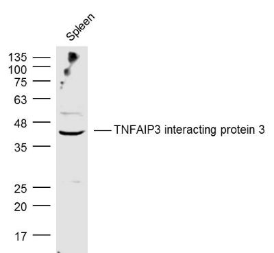
TNFAIP3 interacting protein 3 Polyclonal Antibody
Purified Rabbit Polyclonal Antibody (Pab)
- SPECIFICATION
- CITATIONS
- PROTOCOLS
- BACKGROUND

Application
| WB, IHC-P, IHC-F, IF, E |
|---|---|
| Primary Accession | Q96KP6 |
| Reactivity | Rat, Bovine |
| Host | Rabbit |
| Clonality | Polyclonal |
| Calculated MW | 39 KDa |
| Physical State | Liquid |
| Immunogen | KLH conjugated synthetic peptide derived from human TNFAIP3 interacting protein 3/ABIN3 |
| Epitope Specificity | 51-150/325 |
| Isotype | IgG |
| Purity | affinity purified by Protein A |
| Buffer | 0.01M TBS (pH7.4) with 1% BSA, 0.02% Proclin300 and 50% Glycerol. |
| SUBUNIT | Interacts with TNFAIP3. Interacts with polyubiquitin. |
| Important Note | This product as supplied is intended for research use only, not for use in human, therapeutic or diagnostic applications. |
| Background Descriptions | ABIN-3 is a member of the A20-binding inhibitor of NF-kappaB activation (ABIN) protein family. Similar to the previously characterized human ABINs (ABIN-1 and ABIN-2), ABIN-3 can bind to A20 and inhibit NF-kappaB activation. In contrast, mouse ABIN-3 is incapable of inhibiting NF-kappaB activation by proinflammatory stimuli because the protein lacks a complete ABIN homology domain, which is required for the funcitonal activity of human ABIN-3. |
| Gene ID | 79931 |
|---|---|
| Other Names | TNFAIP3-interacting protein 3, A20-binding inhibitor of NF-kappa-B activation 3, ABIN-3, Listeria-induced gene protein, TNIP3 (HGNC:19315), ABIN3, LIND |
| Target/Specificity | Highly expressed in lung, lymph node, thymus and fetal liver. Expressed at lower levels in bone marrow, brain, kidney, spleen, leukocytes and tonsils. Could be detected in heart, salivary gland, adrenal gland, pancreas, ovary and fetal brain. High levels detected in liver, colon, small intestine, muscle, stomach, testis, placenta, thyroid, uterus, prostate, skin and PBL. |
| Dilution | WB=1:500-2000,IHC-P=1:100-500,IHC-F=1:100-500,IF=1:100-500,ELISA=1:5000-10000 |
| Storage | Store at -20 ℃ for one year. Avoid repeated freeze/thaw cycles. When reconstituted in sterile pH 7.4 0.01M PBS or diluent of antibody the antibody is stable for at least two weeks at 2-4 ℃. |
| Name | TNIP3 (HGNC:19315) |
|---|---|
| Synonyms | ABIN3, LIND |
| Function | Binds to zinc finger protein TNFAIP3 and inhibits NF-kappa-B activation induced by tumor necrosis factor, Toll-like receptor 4 (TLR4), interleukin-1 and 12-O-tetradecanoylphorbol-13-acetate. Overexpression inhibits NF-kappa-B-dependent gene expression in response to lipopolysaccharide at a level downstream of TRAF6 and upstream of IKBKB. NF-kappa-B inhibition is independent of TNFAIP3 binding. |
| Tissue Location | Highly expressed in lung, lymph node, thymus and fetal liver. Expressed at lower levels in bone marrow, brain, kidney, spleen, leukocytes and tonsils. Could be detected in heart, salivary gland, adrenal gland, pancreas, ovary and fetal brain. High levels detected in liver, colon, small intestine, muscle, stomach, testis, placenta, thyroid, uterus, prostate, skin and PBL |
Research Areas
Citations (0)

Thousands of laboratories across the world have published research that depended on the performance of antibodies from Abcepta to advance their research. Check out links to articles that cite our products in major peer-reviewed journals, organized by research category.
Submit your citation using an Abcepta antibody to
info@abcepta.com, and receive a free "I Love Antibodies" mug.
info@abcepta.com, and receive a free "I Love Antibodies" mug.

Application Protocols
Provided below are standard protocols that you may find useful for product applications.
Abcepta welcomes feedback from its customers.
If you have used an Abcepta product and would like to share how it has performed, please click on the "Submit Review" button and provide the requested information. Our staff will examine and post your review and contact you if needed.
If you have any additional inquiries please email technical services at tech@abcepta.com.
$ 385.00
Cat# AP58679
Ordering Information
United States
AlbaniaAustraliaAustriaBelgiumBosnia & HerzegovinaBrazilBulgariaCanadaCentral AmericaChinaCroatiaCyprusCzech RepublicDenmarkEstoniaFinlandFranceGermanyGreeceHong KongHungaryIcelandIndiaIndonesiaIrelandIsraelItalyJapanLatviaLithuaniaLuxembourgMacedoniaMalaysiaMaltaMexicoNetherlandsNew ZealandNorwayPakistanPolandPortugalRomaniaSerbiaSingaporeSlovakiaSloveniaSouth AfricaSouth KoreaSpainSwedenSwitzerlandTaiwanTurkeyUnited KingdomUnited StatesVietnamWorldwideOthers
USA Headquarters
(888) 735-7227 / (858) 622-0099 or (858) 875-1900
Other Products
Shipping Information
Domestic orders (in stock items)
Shipped out the same day. Orders placed after 1 PM (PST) will ship out the next business day.
International orders
Contact your local distributors
