


 Foundational characteristics of cancer include proliferation, angiogenesis, migration, evasion of apoptosis, and cellular immortality. Find key markers for these cellular processes and antibodies to detect them.
 The SUMOplot™ Analysis Program predicts and scores sumoylation sites in your protein. SUMOylation is a post-translational modification involved in various cellular processes, such as nuclear-cytosolic transport, transcriptional regulation, apoptosis, protein stability, response to stress, and progression through the cell cycle.
 The Autophagy Receptor Motif Plotter predicts and scores autophagy receptor binding sites in your protein. Identifying proteins connected to this pathway is critical to understanding the role of autophagy in physiological as well as pathological processes such as development, differentiation, neurodegenerative diseases, stress, infection, and cancer.
> home > Products > Primary Antibodies > Antibody Collections > Fc gamma R-mediated phagocytosis > NCF1 Antibody (N-term)
NCF1 Antibody (N-term)
Affinity Purified Rabbit Polyclonal Antibody (Pab)
- SPECIFICATION
- CITATIONS
- PROTOCOLS
- BACKGROUND

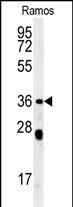
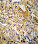
Application
| WB, IHC-P, E |
|---|---|
| Primary Accession | P14598 |
| Other Accession | O77774, A8MVU1, A6NI72, NP_000256.3 |
| Reactivity | Human |
| Predicted | Bovine |
| Host | Rabbit |
| Clonality | Polyclonal |
| Isotype | Rabbit IgG |
| Calculated MW | 44682 Da |
| Antigen Region | 26-52 aa |
| Gene ID | 653361 |
|---|---|
| Other Names | Neutrophil cytosol factor 1, NCF-1, 47 kDa autosomal chronic granulomatous disease protein, 47 kDa neutrophil oxidase factor, NCF-47K, Neutrophil NADPH oxidase factor 1, Nox organizer 2, Nox-organizing protein 2, SH3 and PX domain-containing protein 1A, p47-phox, NCF1, NOXO2, SH3PXD1A |
| Target/Specificity | This NCF1 antibody is generated from rabbits immunized with a KLH conjugated synthetic peptide between 26-52 amino acids from the N-terminal region of human NCF1. |
| Dilution | WB~~1:1000 IHC-P~~1:50~100 E~~Use at an assay dependent concentration. |
| Format | Purified polyclonal antibody supplied in PBS with 0.09% (W/V) sodium azide. This antibody is purified through a protein A column, followed by peptide affinity purification. |
| Storage | Maintain refrigerated at 2-8°C for up to 2 weeks. For long term storage store at -20°C in small aliquots to prevent freeze-thaw cycles. |
| Precautions | NCF1 Antibody (N-term) is for research use only and not for use in diagnostic or therapeutic procedures. |
| Name | NCF1 (HGNC:7660) |
|---|---|
| Synonyms | NOXO2, SH3PXD1A |
| Function | Subunit of the phagocyte NADPH oxidase complex that mediates the transfer of electrons from cytosolic NADPH to O2 to produce the superoxide anion (O2(-)) (PubMed:2547247, PubMed:2550933, PubMed:38355798). In the activated complex, electrons are first transferred from NADPH to flavin adenine dinucleotide (FAD) and subsequently transferred via two heme molecules to molecular oxygen, producing superoxide through an outer-sphere reaction (PubMed:38355798). Activation of the NADPH oxidase complex is initiated by the assembly of cytosolic subunits of the NADPH oxidase complex with the core NADPH oxidase complex to form a complex at the plasma membrane or phagosomal membrane (PubMed:38355798). This activation process is initiated by phosphorylation dependent binding of the cytosolic NCF1/p47-phox subunit to the C-terminus of CYBA/p22-phox (PubMed:12732142, PubMed:19801500). |
| Cellular Location | Cytoplasm, cytosol. Membrane; Peripheral membrane protein; Cytoplasmic side |
| Tissue Location | Detected in peripheral blood monocytes and neutrophils (at protein level). |
Research Areas
Citations (0)

Thousands of laboratories across the world have published research that depended on the performance of antibodies from Abcepta to advance their research. Check out links to articles that cite our products in major peer-reviewed journals, organized by research category.
Submit your citation using an Abcepta antibody to
info@abcepta.com, and receive a free "I Love Antibodies" mug.
info@abcepta.com, and receive a free "I Love Antibodies" mug.

Application Protocols
Provided below are standard protocols that you may find useful for product applications.
Abcepta welcomes feedback from its customers.
If you have used an Abcepta product and would like to share how it has performed, please click on the "Submit Review" button and provide the requested information. Our staff will examine and post your review and contact you if needed.
If you have any additional inquiries please email technical services at tech@abcepta.com.
$ 150.00
$ 385.00
Cat# AP5882a
Ordering Information
United States
AlbaniaAustraliaAustriaBelgiumBosnia & HerzegovinaBrazilBulgariaCanadaCentral AmericaChinaCroatiaCyprusCzech RepublicDenmarkEstoniaFinlandFranceGermanyGreeceHong KongHungaryIcelandIndiaIndonesiaIrelandIsraelItalyJapanLatviaLithuaniaLuxembourgMacedoniaMalaysiaMaltaMexicoNetherlandsNew ZealandNorwayPakistanPolandPortugalRomaniaSerbiaSingaporeSlovakiaSloveniaSouth AfricaSouth KoreaSpainSwedenSwitzerlandTaiwanTurkeyUnited KingdomUnited StatesVietnamWorldwideOthers
USA Headquarters
(888) 735-7227 / (858) 622-0099 or (858) 875-1900
Other Products
Shipping Information
Domestic orders (in stock items)
Shipped out the same day. Orders placed after 1 PM (PST) will ship out the next business day.
International orders
Contact your local distributors
