


 Foundational characteristics of cancer include proliferation, angiogenesis, migration, evasion of apoptosis, and cellular immortality. Find key markers for these cellular processes and antibodies to detect them.
 The SUMOplot™ Analysis Program predicts and scores sumoylation sites in your protein. SUMOylation is a post-translational modification involved in various cellular processes, such as nuclear-cytosolic transport, transcriptional regulation, apoptosis, protein stability, response to stress, and progression through the cell cycle.
 The Autophagy Receptor Motif Plotter predicts and scores autophagy receptor binding sites in your protein. Identifying proteins connected to this pathway is critical to understanding the role of autophagy in physiological as well as pathological processes such as development, differentiation, neurodegenerative diseases, stress, infection, and cancer.
RDH13 Polyclonal Antibody
Purified Rabbit Polyclonal Antibody (Pab)
- SPECIFICATION
- CITATIONS
- PROTOCOLS
- BACKGROUND

Application
| WB, IHC-P, IHC-F, IF, E |
|---|---|
| Primary Accession | Q8NBN7 |
| Reactivity | Rat, Dog, Bovine |
| Host | Rabbit |
| Clonality | Polyclonal |
| Calculated MW | 36 KDa |
| Physical State | Liquid |
| Immunogen | KLH conjugated synthetic peptide derived from human RDH13 |
| Epitope Specificity | 101-200/331 |
| Isotype | IgG |
| Purity | affinity purified by Protein A |
| Buffer | 0.01M TBS (pH7.4) with 1% BSA, 0.02% Proclin300 and 50% Glycerol. |
| SIMILARITY | Belongs to the short-chain dehydrogenases/reductases (SDR) family. |
| Important Note | This product as supplied is intended for research use only, not for use in human, therapeutic or diagnostic applications. |
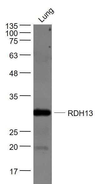
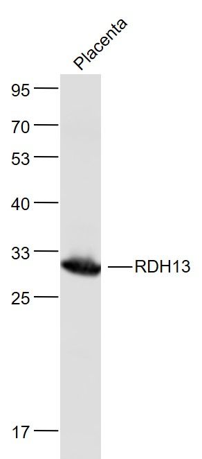
| Background Descriptions | RDH13, also known as all-trans and 9-cis retinol dehydrogenase 13 or SDR7C3, is a 331 amino acid mitochondrial protein belonging to the short-chain dehydrogenases/reductases (SDR) family. Widely expressed, mostly in eye, pancreas, placenta and lung, RDH13 localizes on the outer side of the inner mitochondrial membrane. Related to microsomal retinoid oxidoreductase RDH11, RDH13 is considered to be a major enzyme among the RDH family of proteins. Catalytically active, RDH13 recognizes retinoids as substrates and may function in retinoic acid production. RDH13 may function to protect the mitochondria against oxidative stress. Leber congenital amaurosis (LCA) type 3, an inherited autosomal recessive retinal disease, has been associated with defects of RDH13. LCA represents the most common genetic cause of congenital visual impairment in infants and children. |
| Gene ID | 112724 |
|---|---|
| Other Names | Retinol dehydrogenase 13, 1.1.1.300, Short chain dehydrogenase/reductase family 7C member 3, RDH13, SDR7C3 |
| Target/Specificity | Expressed mostly in eye, pancreas, placenta and lung. In the retina, detected in the inner segment of the photoreceptor cells. Weak signals were observed in a small population of inner nuclear neurons and the inner plexiform layer. |
| Dilution | WB=1:500-2000,IHC-P=1:100-500,IHC-F=1:100-500,IF=1:50-200,ELISA=1:5000-10000 |
| Storage | Store at -20 ℃ for one year. Avoid repeated freeze/thaw cycles. When reconstituted in sterile pH 7.4 0.01M PBS or diluent of antibody the antibody is stable for at least two weeks at 2-4 ℃. |
| Name | RDH13 |
|---|---|
| Synonyms | SDR7C3 |
| Function | Retinol dehydrogenase with a clear preference for NADP. Oxidizes all-trans-retinol, but seems to reduce all-trans-retinal with much higher efficiency (PubMed:18039331). Has no activity toward steroids (PubMed:18039331). |
| Cellular Location | Mitochondrion inner membrane; Peripheral membrane protein. Note=Localized on the outer side of the inner mitochondrial membrane. |
| Tissue Location | Widely expressed (PubMed:18039331). In the retina, detected in the inner segment of the photoreceptor cells. Weak signals are observed in a small population of inner nuclear neurons and the inner plexiform layer (PubMed:12226107). |
Research Areas
Citations (0)

Thousands of laboratories across the world have published research that depended on the performance of antibodies from Abcepta to advance their research. Check out links to articles that cite our products in major peer-reviewed journals, organized by research category.
Submit your citation using an Abcepta antibody to
info@abcepta.com, and receive a free "I Love Antibodies" mug.
info@abcepta.com, and receive a free "I Love Antibodies" mug.

Application Protocols
Provided below are standard protocols that you may find useful for product applications.
Abcepta welcomes feedback from its customers.
If you have used an Abcepta product and would like to share how it has performed, please click on the "Submit Review" button and provide the requested information. Our staff will examine and post your review and contact you if needed.
If you have any additional inquiries please email technical services at tech@abcepta.com.
$ 385.00
Cat# AP58968
Ordering Information
United States
AlbaniaAustraliaAustriaBelgiumBosnia & HerzegovinaBrazilBulgariaCanadaCentral AmericaChinaCroatiaCyprusCzech RepublicDenmarkEstoniaFinlandFranceGermanyGreeceHong KongHungaryIcelandIndiaIndonesiaIrelandIsraelItalyJapanLatviaLithuaniaLuxembourgMacedoniaMalaysiaMaltaMexicoNetherlandsNew ZealandNorwayPakistanPolandPortugalRomaniaSerbiaSingaporeSlovakiaSloveniaSouth AfricaSouth KoreaSpainSwedenSwitzerlandTaiwanTurkeyUnited KingdomUnited StatesVietnamWorldwideOthers
USA Headquarters
(888) 735-7227 / (858) 622-0099 or (858) 875-1900
Shipping Information
Domestic orders (in stock items)
Shipped out the same day. Orders placed after 1 PM (PST) will ship out the next business day.
International orders
Contact your local distributors
