


 Foundational characteristics of cancer include proliferation, angiogenesis, migration, evasion of apoptosis, and cellular immortality. Find key markers for these cellular processes and antibodies to detect them.
 The SUMOplot™ Analysis Program predicts and scores sumoylation sites in your protein. SUMOylation is a post-translational modification involved in various cellular processes, such as nuclear-cytosolic transport, transcriptional regulation, apoptosis, protein stability, response to stress, and progression through the cell cycle.
 The Autophagy Receptor Motif Plotter predicts and scores autophagy receptor binding sites in your protein. Identifying proteins connected to this pathway is critical to understanding the role of autophagy in physiological as well as pathological processes such as development, differentiation, neurodegenerative diseases, stress, infection, and cancer.
> home > Products > Primary Antibodies > Antibody Collections > Prostate negative > NEU1/Neuraminidase Polyclonal Antibody
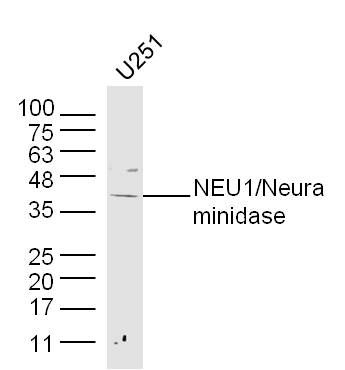
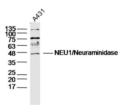
NEU1/Neuraminidase Polyclonal Antibody
Purified Rabbit Polyclonal Antibody (Pab)
- SPECIFICATION
- CITATIONS
- PROTOCOLS
- BACKGROUND

Application
| WB, IHC-P, IHC-F, IF, ICC, E |
|---|---|
| Primary Accession | Q99519 |
| Reactivity | Rat, Bovine |
| Host | Rabbit |
| Clonality | Polyclonal |
| Calculated MW | 45 KDa |
| Physical State | Liquid |
| Immunogen | KLH conjugated synthetic peptide derived from human NEU1/Neuraminidase |
| Epitope Specificity | 151-250/415 |
| Isotype | IgG |
| Purity | affinity purified by Protein A |
| Buffer | 0.01M TBS (pH7.4) with 1% BSA, 0.02% Proclin300 and 50% Glycerol. |
| SUBCELLULAR LOCATION | Lysosome membrane. Lysosome lumen. Cell membrane. Cytoplasmic vesicle. Localized not only on the inner side of the lysosomal membrane and in the lysosomal lumen, but also on the plasma membrane and in intracellular vesicles. |
| SIMILARITY | Belongs to the glycosyl hydrolase 33 family. Contains 4 BNR repeats. |
| SUBUNIT | Interacts with cathepsin A (protective protein),beta-galactosidase and N-acetylgalactosamine-6-sulfate sulfatase in a multienzyme complex. |
| Post-translational modifications | N-glycosylated. Phosphorylation of tyrosine within the internalization signal results in inhibition of sialidase internalization and blockage on the plasma membrane. |
| DISEASE | Defects in NEU1 are the cause of sialidosis (SIALIDOSIS) [MIM:256550]. It is a lysosomal storage disease occurring as two types with various manifestations. Type 1 sialidosis (cherry red spot-myoclonus syndrome or normosomatic type) is late-onset and it is characterized by the formation of cherry red macular spots in childhood, progressive debilitating myoclonus, insiduous visual loss and rarely ataxia. The diagnosis can be confirmed by the screening of the urine for sialyloligosaccharides. Type 2 sialidosis (also known as dysmorphic type) occurs as several variants of increasing severity with earlier age of onset. It is characterized by the presence of abnormal somatic features including coarse facies and dysostosis multiplex, vertebral deformities, mental retardation, cherry-red spot/myoclonus, sialuria, cytoplasmic vacuolation of peripheral lymphocytes, bone marrow cells and conjunctival epithelial cells. |
| Important Note | This product as supplied is intended for research use only, not for use in human, therapeutic or diagnostic applications. |
| Background Descriptions | The protein encoded by this gene is a lysosomal enzyme that cleaves terminal sialic acid residues from substrates such as glycoproteins and glycolipids. In the lysosome, this enzyme is part of a heterotrimeric complex together with beta-galactosidase and cathepsin A (the latter is also referred to as 'protective protein'). Mutations in this gene can lead to sialidosis, a lysosomal storage disease that can be type 1 (cherry red spot-myoclonus syndrome or normosomatic type), which is late-onset, or type 2 (the dysmorphic type), which occurs at an earlier age with increased severity. [provided by RefSeq, Jul 2008] |
| Gene ID | 4758 |
|---|---|
| Other Names | Sialidase-1, 3.2.1.18, Acetylneuraminyl hydrolase, G9 sialidase, Lysosomal sialidase, N-acetyl-alpha-neuraminidase 1, NEU1, NANH |
| Target/Specificity | Highly expressed in pancreas, followed by skeletal muscle, kidney, placenta, heart, lung and liver. Weakly expressed in brain. |
| Dilution | WB=1:500-2000,IHC-P=1:100-500,IHC-F=1:100-500,ICC=1:100-500,IF=1:100-500,ELISA=1:5000-10000 |
| Storage | Store at -20 ℃ for one year. Avoid repeated freeze/thaw cycles. When reconstituted in sterile pH 7.4 0.01M PBS or diluent of antibody the antibody is stable for at least two weeks at 2-4 ℃. |
| Name | NEU1 |
|---|---|
| Synonyms | NANH |
| Function | Catalyzes the removal of sialic acid (N-acetylneuraminic acid) moieties from glycoproteins and glycolipids. To be active, it is strictly dependent on its presence in the multienzyme complex. Appears to have a preference for alpha 2-3 and alpha 2-6 sialyl linkage. |
| Cellular Location | Lysosome membrane; Peripheral membrane protein; Lumenal side. Lysosome lumen. Cell membrane. Cytoplasmic vesicle Lysosome. Note=Localized not only on the inner side of the lysosomal membrane and in the lysosomal lumen, but also on the plasma membrane and in intracellular vesicles |
| Tissue Location | Highly expressed in pancreas, followed by skeletal muscle, kidney, placenta, heart, lung and liver. Weakly expressed in brain. |
Research Areas
Citations (0)

Thousands of laboratories across the world have published research that depended on the performance of antibodies from Abcepta to advance their research. Check out links to articles that cite our products in major peer-reviewed journals, organized by research category.
Submit your citation using an Abcepta antibody to
info@abcepta.com, and receive a free "I Love Antibodies" mug.
info@abcepta.com, and receive a free "I Love Antibodies" mug.

Application Protocols
Provided below are standard protocols that you may find useful for product applications.
Abcepta welcomes feedback from its customers.
If you have used an Abcepta product and would like to share how it has performed, please click on the "Submit Review" button and provide the requested information. Our staff will examine and post your review and contact you if needed.
If you have any additional inquiries please email technical services at tech@abcepta.com.
$ 385.00
Cat# AP59051
Ordering Information
United States
AlbaniaAustraliaAustriaBelgiumBosnia & HerzegovinaBrazilBulgariaCanadaCentral AmericaChinaCroatiaCyprusCzech RepublicDenmarkEstoniaFinlandFranceGermanyGreeceHong KongHungaryIcelandIndiaIndonesiaIrelandIsraelItalyJapanLatviaLithuaniaLuxembourgMacedoniaMalaysiaMaltaMexicoNetherlandsNew ZealandNorwayPakistanPolandPortugalRomaniaSerbiaSingaporeSlovakiaSloveniaSouth AfricaSouth KoreaSpainSwedenSwitzerlandTaiwanTurkeyUnited KingdomUnited StatesVietnamWorldwideOthers
USA Headquarters
(888) 735-7227 / (858) 622-0099 or (858) 875-1900
Other Products
Shipping Information
Domestic orders (in stock items)
Shipped out the same day. Orders placed after 1 PM (PST) will ship out the next business day.
International orders
Contact your local distributors
