


 Foundational characteristics of cancer include proliferation, angiogenesis, migration, evasion of apoptosis, and cellular immortality. Find key markers for these cellular processes and antibodies to detect them.
 The SUMOplot™ Analysis Program predicts and scores sumoylation sites in your protein. SUMOylation is a post-translational modification involved in various cellular processes, such as nuclear-cytosolic transport, transcriptional regulation, apoptosis, protein stability, response to stress, and progression through the cell cycle.
 The Autophagy Receptor Motif Plotter predicts and scores autophagy receptor binding sites in your protein. Identifying proteins connected to this pathway is critical to understanding the role of autophagy in physiological as well as pathological processes such as development, differentiation, neurodegenerative diseases, stress, infection, and cancer.
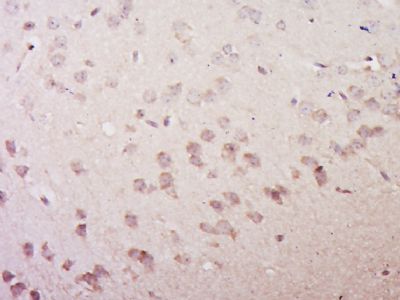
VGLUT3/SLC17A8 Polyclonal Antibody
Purified Rabbit Polyclonal Antibody (Pab)
- SPECIFICATION
- CITATIONS
- PROTOCOLS
- BACKGROUND

Application
| IHC-P, IHC-F, IF, ICC, E |
|---|---|
| Primary Accession | Q8NDX2 |
| Reactivity | Rat, Dog, Bovine |
| Host | Rabbit |
| Clonality | Polyclonal |
| Calculated MW | 65 KDa |
| Physical State | Liquid |
| Immunogen | KLH conjugated synthetic peptide derived from human VGLUT3/SLC17A8 |
| Epitope Specificity | 1-100/589 |
| Isotype | IgG |
| Purity | affinity purified by Protein A |
| Buffer | Preservative: 0.02% Proclin300, Constituents: 1% BSA, 0.01M PBS, pH7.4. |
| SUBCELLULAR LOCATION | Cytoplasmic vesicle > secretory vesicle > synaptic vesicle membrane. Membrane. Cell junction > synapse > synaptosome. |
| SIMILARITY | Belongs to the major facilitator superfamily. Sodium/anion cotransporter family. VGLUT subfamily. |
| DISEASE | Defects in SLC17A8 are the cause of deafness autosomal dominant type 25 (DFNA25) [MIM:605583]. DFNA25 is a form of sensorineural hearing loss. The expression of DFNA25 deafness is variable in terms of onset and rate of progression, with an age-dependent penetrance resembling an early-onset presbycusis, or senile deafness, a progressive bilateral loss of hearing that occurs in the aged. |
| Important Note | This product as supplied is intended for research use only, not for use in human, therapeutic or diagnostic applications. |
| Background Descriptions | This gene encodes a vesicular glutamate transporter. The encoded protein transports the neurotransmitter glutamate into synaptic vesicles before it is released into the synaptic cleft. Mutations in this gene are the cause of autosomal-dominant nonsyndromic type 25 deafness. Alternate splicing results in multiple transcript variants.[provided by RefSeq, May 2010] |
| Gene ID | 246213 |
|---|---|
| Other Names | Vesicular glutamate transporter 3, VGluT3, Solute carrier family 17 member 8, SLC17A8, VGLUT3 |
| Target/Specificity | Expressed in amygdala, cerebellum, hippocampus, medulla, spinal cord and thalamus. |
| Dilution | IHC-P=1:100-500,IHC-F=1:100-500,ICC=1:100-500,IF=1:100-500,ELISA=1:5000-10000 |
| Storage | Store at -20 ℃ for one year. Avoid repeated freeze/thaw cycles. When reconstituted in sterile pH 7.4 0.01M PBS or diluent of antibody the antibody is stable for at least two weeks at 2-4 ℃. |
| Name | SLC17A8 (HGNC:20151) |
|---|---|
| Synonyms | VGLUT3 |
| Function | Multifunctional transporter that transports L-glutamate as well as multiple ions such as chloride, sodium and phosphate (PubMed:12151341, PubMed:33440152). At the synaptic vesicle membrane, mainly functions as an uniporter that mediates the uptake of L- glutamate into synaptic vesicles at presynaptic nerve terminals of excitatory neural cells (PubMed:12151341). The L-glutamate uniporter activity is electrogenic and is driven by the proton electrochemical gradient, mainly by the electrical gradient established by the vacuolar H(+)-ATPase across the synaptic vesicle membrane (PubMed:12151341). In addition, functions as a chloride channel that allows a chloride permeation through the synaptic vesicle membrane that affects the proton electrochemical gradient and promotes synaptic vesicles acidification (By similarity). At the plasma membrane, following exocytosis, functions as a symporter of Na(+) and phosphate from the extracellular space to the cytoplasm allowing synaptic phosphate homeostasis regulation (Probable). The symporter activity is electrogenic (PubMed:33440152). Moreover, operates synergistically with SLC18A3/VACHT under a constant H(+) gradient, thereby allowing striatal vesicular acetylcholine uptake (By similarity). |
| Cellular Location | Cytoplasmic vesicle, secretory vesicle, synaptic vesicle membrane {ECO:0000250|UniProtKB:Q7TSF2}. Cell membrane; Multi-pass membrane protein. Synapse, synaptosome {ECO:0000250|UniProtKB:Q7TSF2} |
| Tissue Location | Expressed in amygdala, cerebellum, hippocampus, medulla, spinal cord and thalamus. |
Research Areas
Citations (0)

Thousands of laboratories across the world have published research that depended on the performance of antibodies from Abcepta to advance their research. Check out links to articles that cite our products in major peer-reviewed journals, organized by research category.
Submit your citation using an Abcepta antibody to
info@abcepta.com, and receive a free "I Love Antibodies" mug.
info@abcepta.com, and receive a free "I Love Antibodies" mug.

Application Protocols
Provided below are standard protocols that you may find useful for product applications.
Abcepta welcomes feedback from its customers.
If you have used an Abcepta product and would like to share how it has performed, please click on the "Submit Review" button and provide the requested information. Our staff will examine and post your review and contact you if needed.
If you have any additional inquiries please email technical services at tech@abcepta.com.
$ 385.00
Cat# AP59066
Ordering Information
United States
AlbaniaAustraliaAustriaBelgiumBosnia & HerzegovinaBrazilBulgariaCanadaCentral AmericaChinaCroatiaCyprusCzech RepublicDenmarkEstoniaFinlandFranceGermanyGreeceHong KongHungaryIcelandIndiaIndonesiaIrelandIsraelItalyJapanLatviaLithuaniaLuxembourgMacedoniaMalaysiaMaltaMexicoNetherlandsNew ZealandNorwayPakistanPolandPortugalRomaniaSerbiaSingaporeSlovakiaSloveniaSouth AfricaSouth KoreaSpainSwedenSwitzerlandTaiwanTurkeyUnited KingdomUnited StatesVietnamWorldwideOthers
USA Headquarters
(888) 735-7227 / (858) 622-0099 or (858) 875-1900
Other Products
Shipping Information
Domestic orders (in stock items)
Shipped out the same day. Orders placed after 1 PM (PST) will ship out the next business day.
International orders
Contact your local distributors
