


 Foundational characteristics of cancer include proliferation, angiogenesis, migration, evasion of apoptosis, and cellular immortality. Find key markers for these cellular processes and antibodies to detect them.
 The SUMOplot™ Analysis Program predicts and scores sumoylation sites in your protein. SUMOylation is a post-translational modification involved in various cellular processes, such as nuclear-cytosolic transport, transcriptional regulation, apoptosis, protein stability, response to stress, and progression through the cell cycle.
 The Autophagy Receptor Motif Plotter predicts and scores autophagy receptor binding sites in your protein. Identifying proteins connected to this pathway is critical to understanding the role of autophagy in physiological as well as pathological processes such as development, differentiation, neurodegenerative diseases, stress, infection, and cancer.
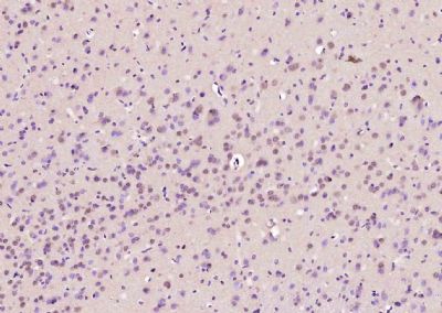
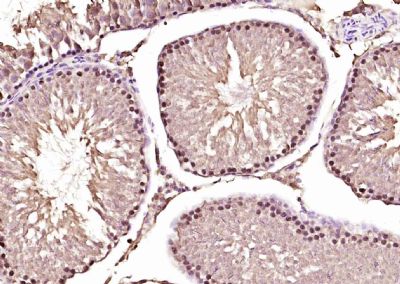
AKIRIN1 Polyclonal Antibody
Purified Rabbit Polyclonal Antibody (Pab)
- SPECIFICATION
- CITATIONS
- PROTOCOLS
- BACKGROUND

Application
| IHC-P, IHC-F, IF, E |
|---|---|
| Primary Accession | Q9H9L7 |
| Reactivity | Rat, Dog |
| Host | Rabbit |
| Clonality | Polyclonal |
| Calculated MW | 22 KDa |
| Physical State | Liquid |
| Immunogen | KLH conjugated synthetic peptide derived from human AKIRIN1 |
| Epitope Specificity | 101-192/192 |
| Isotype | IgG |
| Purity | affinity purified by Protein A |
| Buffer | 0.01M TBS (pH7.4) with 1% BSA, 0.02% Proclin300 and 50% Glycerol. |
| SUBCELLULAR LOCATION | Nuclear |
| SIMILARITY | Belongs to the akirin family. |
| Important Note | This product as supplied is intended for research use only, not for use in human, therapeutic or diagnostic applications. |
| Background Descriptions | AKIRIN1 is dispensable in the mouse, and neither knockout mice nor cells derived from them have obvious distinctive phenotypes. In contrast, Akirin2 is required for development in the mouse and knockout of both Akirin homologs in mice show that Akirin2 is required downstream of toll-like receptor (TLR), TNF-alpha and IL-1beta signaling, and for the production of IL-6. Akirin2 is functionally closer to the single gene in Drosophila, as the homozygous null D. melanogaster Akirin mutants show a similar, mid-to-early embryonic death.The highly conserved, nuclear-localized AKIRIN1 and Akirin2 proteins critically regulate the transcription of NF-kB dependent genes and are required for defense against Gram-negative bacteria in the immune deficiency and NF-kB pathways. |
| Gene ID | 79647 |
|---|---|
| Other Names | Akirin-1, AKIRIN1 (HGNC:25744) |
| Target/Specificity | Widely expressed with the highest expression in heart, liver, placenta and peripheral blood leukocytes. |
| Dilution | IHC-P=1:100-500,IHC-F=1:100-500,IF=1:50-200,ELISA=1:5000-10000 |
| Storage | Store at -20 ℃ for one year. Avoid repeated freeze/thaw cycles. When reconstituted in sterile pH 7.4 0.01M PBS or diluent of antibody the antibody is stable for at least two weeks at 2-4 ℃. |
| Name | AKIRIN1 {ECO:0000303|PubMed:18066067, ECO:0000312|HGNC:HGNC:25744} |
|---|---|
| Function | Molecular adapter that acts as a bridge between proteins, and which is involved skeletal muscle development (By similarity). Functions as a signal transducer for MSTN during skeletal muscle regeneration and myogenesis (By similarity). May regulate chemotaxis of both macrophages and myoblasts by reorganising actin cytoskeleton, leading to more efficient lamellipodia formation via a PI3 kinase dependent pathway (By similarity). In contrast to AKIRIN2, not involved in nuclear import of proteasomes (PubMed:34711951). |
| Cellular Location | Nucleus. |
| Tissue Location | Widely expressed with the highest expression in heart, liver, placenta and peripheral blood leukocytes |
Citations (0)

Thousands of laboratories across the world have published research that depended on the performance of antibodies from Abcepta to advance their research. Check out links to articles that cite our products in major peer-reviewed journals, organized by research category.
Submit your citation using an Abcepta antibody to
info@abcepta.com, and receive a free "I Love Antibodies" mug.
info@abcepta.com, and receive a free "I Love Antibodies" mug.

Application Protocols
Provided below are standard protocols that you may find useful for product applications.
Abcepta welcomes feedback from its customers.
If you have used an Abcepta product and would like to share how it has performed, please click on the "Submit Review" button and provide the requested information. Our staff will examine and post your review and contact you if needed.
If you have any additional inquiries please email technical services at tech@abcepta.com.
$ 385.00
Cat# AP59134
Ordering Information
United States
AlbaniaAustraliaAustriaBelgiumBosnia & HerzegovinaBrazilBulgariaCanadaCentral AmericaChinaCroatiaCyprusCzech RepublicDenmarkEstoniaFinlandFranceGermanyGreeceHong KongHungaryIcelandIndiaIndonesiaIrelandIsraelItalyJapanLatviaLithuaniaLuxembourgMacedoniaMalaysiaMaltaMexicoNetherlandsNew ZealandNorwayPakistanPolandPortugalRomaniaSerbiaSingaporeSlovakiaSloveniaSouth AfricaSouth KoreaSpainSwedenSwitzerlandTaiwanTurkeyUnited KingdomUnited StatesVietnamWorldwideOthers
USA Headquarters
(888) 735-7227 / (858) 622-0099 or (858) 875-1900
Other Products
Shipping Information
Domestic orders (in stock items)
Shipped out the same day. Orders placed after 1 PM (PST) will ship out the next business day.
International orders
Contact your local distributors
