


 Foundational characteristics of cancer include proliferation, angiogenesis, migration, evasion of apoptosis, and cellular immortality. Find key markers for these cellular processes and antibodies to detect them.
 The SUMOplot™ Analysis Program predicts and scores sumoylation sites in your protein. SUMOylation is a post-translational modification involved in various cellular processes, such as nuclear-cytosolic transport, transcriptional regulation, apoptosis, protein stability, response to stress, and progression through the cell cycle.
 The Autophagy Receptor Motif Plotter predicts and scores autophagy receptor binding sites in your protein. Identifying proteins connected to this pathway is critical to understanding the role of autophagy in physiological as well as pathological processes such as development, differentiation, neurodegenerative diseases, stress, infection, and cancer.
AGXT2L2 Polyclonal Antibody
Purified Rabbit Polyclonal Antibody (Pab)
- SPECIFICATION
- CITATIONS
- PROTOCOLS
- BACKGROUND

Application
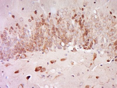
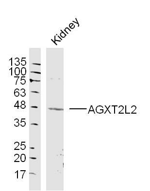
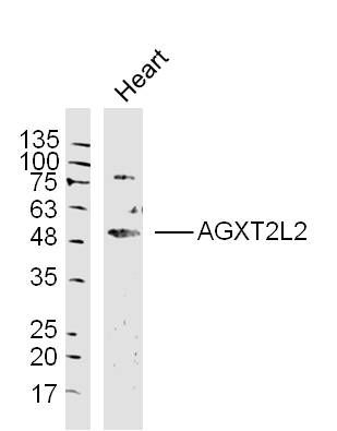
| WB, IHC-P, IHC-F, IF, E |
|---|---|
| Primary Accession | Q8IUZ5 |
| Reactivity | Rat |
| Host | Rabbit |
| Clonality | Polyclonal |
| Calculated MW | 50 KDa |
| Physical State | Liquid |
| Immunogen | KLH conjugated synthetic peptide derived from human AGXT2L2 |
| Epitope Specificity | 381-450/450 |
| Purity | affinity purified by Protein A |
| Buffer | 0.01M TBS (pH7.4) with 1% BSA, 0.02% Proclin300 and 50% Glycerol. |
| SUBCELLULAR LOCATION | Mitochondrial |
| SIMILARITY | Belongs to the class-III pyridoxal-phosphate-dependent aminotransferase family. |
| SUBUNIT | Homotetramer. |
| Important Note | This product as supplied is intended for research use only, not for use in human, therapeutic or diagnostic applications. |
| Background Descriptions | Members of the class-III pyridoxal-phosphate-dependent aminotransferase family, such as AGXT2, catalyze the conversion of glyoxylate to glycine using L-alanine as the amino donor. AGXT2 protects from asymmetric dimethylarginine (ADMA)-induced inhibition in nitric oxide (NO) production. Elevated blood concentrations of ADMA, a methyl derivate of the amino acid arginine and an endogenous inhibitor of nitric oxide (NO) synthase, is produced by the physiological degradation of methylated proteins and is found in association with diabetes, hypertension, congestive heart failure and atherosclerosis. AGXT2L2 (alanine-glyoxylate aminotransferase 2-like 2) is a 450 amino acid pyridoxal phosphate that exists as a homotetramer. Belonging to the class-III pyridoxal-phosphate-dependent aminotransferase family, AGXT2L2 localizes to the mitochondria and exists as three alternatively spliced isoforms. Encoded by a gene located on human chromosome 5q35.3, AGXT2L2 may have similar functions as AGXT2. |
| Gene ID | 85007 |
|---|---|
| Other Names | 5-phosphohydroxy-L-lysine phospho-lyase, 4.2.3.134, Alanine--glyoxylate aminotransferase 2-like 2, PHYKPL, AGXT2L2 |
| Dilution | WB=1:500-2000,IHC-P=1:100-500,IHC-F=1:100-500,IF=1:50-200,ELISA=1:5000-10000 |
| Storage | Store at -20 ℃ for one year. Avoid repeated freeze/thaw cycles. When reconstituted in sterile pH 7.4 0.01M PBS or diluent of antibody the antibody is stable for at least two weeks at 2-4 ℃. |
| Name | PHYKPL |
|---|---|
| Synonyms | AGXT2L2 {ECO:0000303|PubMed:22241472} |
| Function | Catalyzes the pyridoxal-phosphate-dependent breakdown of 5- phosphohydroxy-L-lysine, converting it to ammonia, inorganic phosphate and 2-aminoadipate semialdehyde. |
| Cellular Location | Mitochondrion. |
Research Areas
Citations (0)

Thousands of laboratories across the world have published research that depended on the performance of antibodies from Abcepta to advance their research. Check out links to articles that cite our products in major peer-reviewed journals, organized by research category.
Submit your citation using an Abcepta antibody to
info@abcepta.com, and receive a free "I Love Antibodies" mug.
info@abcepta.com, and receive a free "I Love Antibodies" mug.

Application Protocols
Provided below are standard protocols that you may find useful for product applications.
Abcepta welcomes feedback from its customers.
If you have used an Abcepta product and would like to share how it has performed, please click on the "Submit Review" button and provide the requested information. Our staff will examine and post your review and contact you if needed.
If you have any additional inquiries please email technical services at tech@abcepta.com.
$ 385.00
Cat# AP59140
Ordering Information
United States
AlbaniaAustraliaAustriaBelgiumBosnia & HerzegovinaBrazilBulgariaCanadaCentral AmericaChinaCroatiaCyprusCzech RepublicDenmarkEstoniaFinlandFranceGermanyGreeceHong KongHungaryIcelandIndiaIndonesiaIrelandIsraelItalyJapanLatviaLithuaniaLuxembourgMacedoniaMalaysiaMaltaMexicoNetherlandsNew ZealandNorwayPakistanPolandPortugalRomaniaSerbiaSingaporeSlovakiaSloveniaSouth AfricaSouth KoreaSpainSwedenSwitzerlandTaiwanTurkeyUnited KingdomUnited StatesVietnamWorldwideOthers
USA Headquarters
(888) 735-7227 / (858) 622-0099 or (858) 875-1900
Other Products
Shipping Information
Domestic orders (in stock items)
Shipped out the same day. Orders placed after 1 PM (PST) will ship out the next business day.
International orders
Contact your local distributors
