


 Foundational characteristics of cancer include proliferation, angiogenesis, migration, evasion of apoptosis, and cellular immortality. Find key markers for these cellular processes and antibodies to detect them.
 The SUMOplot™ Analysis Program predicts and scores sumoylation sites in your protein. SUMOylation is a post-translational modification involved in various cellular processes, such as nuclear-cytosolic transport, transcriptional regulation, apoptosis, protein stability, response to stress, and progression through the cell cycle.
 The Autophagy Receptor Motif Plotter predicts and scores autophagy receptor binding sites in your protein. Identifying proteins connected to this pathway is critical to understanding the role of autophagy in physiological as well as pathological processes such as development, differentiation, neurodegenerative diseases, stress, infection, and cancer.
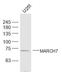
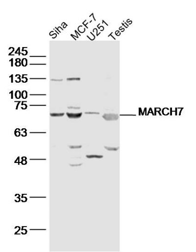
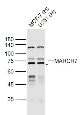
MARCH7 Polyclonal Antibody
Purified Rabbit Polyclonal Antibody (Pab)

- SPECIFICATION
- CITATIONS
- PROTOCOLS
- BACKGROUND

Application
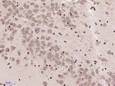
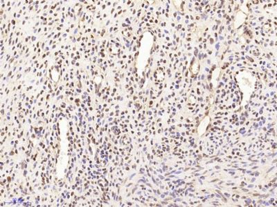
| WB, IHC-P, IHC-F, IF, E |
|---|---|
| Primary Accession | Q9H992 |
| Reactivity | Rat, Pig, Dog, Bovine |
| Host | Rabbit |
| Clonality | Polyclonal |
| Calculated MW | 78 KDa |
| Physical State | Liquid |
| Immunogen | KLH conjugated synthetic peptide derived from human MARCH7/Axotrophin |
| Epitope Specificity | 31-130/704 |
| Isotype | IgG |
| Purity | affinity purified by Protein A |
| Buffer | 0.01M TBS (pH7.4) with 1% BSA, 0.02% Proclin300 and 50% Glycerol. |
| SIMILARITY | Contains 1 RING-CH-type zinc finger. |
| Important Note | This product as supplied is intended for research use only, not for use in human, therapeutic or diagnostic applications. |
| Background Descriptions | Axotrophin is a stem cell gene that encodes a protein which is involved in T lymphocyte regulation (especially in regulating the proliferation) and leukemia inhibitory factor (LIF) release. LIF is a neuropoietic cytokine that is important for stem cell regulation and thymocyte stimulation. Both Axotrophin and LIF are linked to transplantation intolerance. Axotrophin is also involved in corpus callosum differentiation and may play a role in glial cell line-derived neurotrophic factor (GDNF)-dependent sensory neuron survival in the substantia gelatinosa of the adult spinal cord. Axotrophin is primarily expressed in the hippocampus, cortex, purkinje and granule cells of the cerebellum. |
| Gene ID | 64844 |
|---|---|
| Other Names | E3 ubiquitin-protein ligase MARCHF7, 2.3.2.27, Axotrophin, Membrane-associated RING finger protein 7, Membrane-associated RING-CH protein VII, MARCH-VII, RING finger protein 177, RING-type E3 ubiquitin transferase MARCHF7, MARCHF7 (HGNC:17393), AXOT, MARCH7, RNF177 |
| Dilution | WB=1:500-2000,IHC-P=1:100-500,IHC-F=1:100-500,IF=1:50-200,ELISA=1:5000-10000 |
| Storage | Store at -20 ℃ for one year. Avoid repeated freeze/thaw cycles. When reconstituted in sterile pH 7.4 0.01M PBS or diluent of antibody the antibody is stable for at least two weeks at 2-4 ℃. |
| Name | MARCHF7 (HGNC:17393) |
|---|---|
| Synonyms | AXOT, MARCH7, RNF177 |
| Function | E3 ubiquitin-protein ligase which may specifically enhance the E2 activity of HIP2. E3 ubiquitin ligases accept ubiquitin from an E2 ubiquitin-conjugating enzyme in the form of a thioester and then directly transfer the ubiquitin to targeted substrates (PubMed:16868077). May be involved in T-cell proliferation by regulating LIF secretion (By similarity). May play a role in lysosome homeostasis (PubMed:31270356). Promotes 'Lys-6', 'Lys-11' and 'Lys-63'- linked mixed polyubiquitination on ATG14 leading to the inhibition of autophagy by impairing the interaction between ATG14 and STX7 (PubMed:37632749). Participates in the dopamine-mediated negative regulation of the NLRP3 inflammasome by promoting its uibiquitination and subsequent degradation (PubMed:25594175). |
| Cellular Location | Cytoplasm. |
Research Areas
Citations (0)

Thousands of laboratories across the world have published research that depended on the performance of antibodies from Abcepta to advance their research. Check out links to articles that cite our products in major peer-reviewed journals, organized by research category.
Submit your citation using an Abcepta antibody to
info@abcepta.com, and receive a free "I Love Antibodies" mug.
info@abcepta.com, and receive a free "I Love Antibodies" mug.

Application Protocols
Provided below are standard protocols that you may find useful for product applications.
Abcepta welcomes feedback from its customers.
If you have used an Abcepta product and would like to share how it has performed, please click on the "Submit Review" button and provide the requested information. Our staff will examine and post your review and contact you if needed.
If you have any additional inquiries please email technical services at tech@abcepta.com.
$ 385.00
Cat# AP59230
Ordering Information
United States
AlbaniaAustraliaAustriaBelgiumBosnia & HerzegovinaBrazilBulgariaCanadaCentral AmericaChinaCroatiaCyprusCzech RepublicDenmarkEstoniaFinlandFranceGermanyGreeceHong KongHungaryIcelandIndiaIndonesiaIrelandIsraelItalyJapanLatviaLithuaniaLuxembourgMacedoniaMalaysiaMaltaMexicoNetherlandsNew ZealandNorwayPakistanPolandPortugalRomaniaSerbiaSingaporeSlovakiaSloveniaSouth AfricaSouth KoreaSpainSwedenSwitzerlandTaiwanTurkeyUnited KingdomUnited StatesVietnamWorldwideOthers
USA Headquarters
(888) 735-7227 / (858) 622-0099 or (858) 875-1900
Other Products
Shipping Information
Domestic orders (in stock items)
Shipped out the same day. Orders placed after 1 PM (PST) will ship out the next business day.
International orders
Contact your local distributors
