


 Foundational characteristics of cancer include proliferation, angiogenesis, migration, evasion of apoptosis, and cellular immortality. Find key markers for these cellular processes and antibodies to detect them.
 The SUMOplot™ Analysis Program predicts and scores sumoylation sites in your protein. SUMOylation is a post-translational modification involved in various cellular processes, such as nuclear-cytosolic transport, transcriptional regulation, apoptosis, protein stability, response to stress, and progression through the cell cycle.
 The Autophagy Receptor Motif Plotter predicts and scores autophagy receptor binding sites in your protein. Identifying proteins connected to this pathway is critical to understanding the role of autophagy in physiological as well as pathological processes such as development, differentiation, neurodegenerative diseases, stress, infection, and cancer.
SHARPIN Polyclonal Antibody
Purified Rabbit Polyclonal Antibody (Pab)
- SPECIFICATION
- CITATIONS
- PROTOCOLS
- BACKGROUND

Application
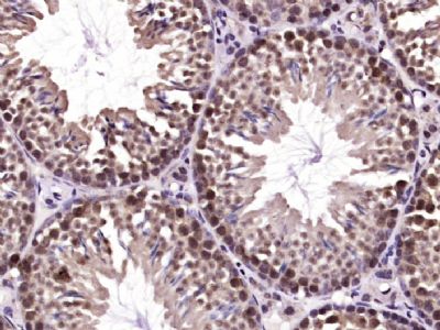
| IHC-P, IHC-F, IF, ICC, E |
|---|---|
| Primary Accession | Q9H0F6 |
| Reactivity | Rat, Dog |
| Host | Rabbit |
| Clonality | Polyclonal |
| Calculated MW | 40 KDa |
| Physical State | Liquid |
| Immunogen | KLH conjugated synthetic peptide derived from human SHARPIN |
| Epitope Specificity | 271-387/387 |
| Isotype | IgG |
| Purity | affinity purified by Protein A |
| Buffer | 0.01M TBS (pH7.4) with 1% BSA, 0.02% Proclin300 and 50% Glycerol. |
| SUBCELLULAR LOCATION | Cytoplasm. Enriched at synaptic sites in mature neurons where it colocalizes with SHANK1. |
| SIMILARITY | Contains 1 RanBP2-type zinc finger. |
| SUBUNIT | Monomer and homodimer. Interacts with SHANK1, EYA1 and EYA2 (By similarity). Component of the LUBAC complex (linear ubiquitin chain assembly complex) which consists of SHARPIN, RBCK1 and RNF31. LUBAC has a MW of approximative 600 kDa suggesting a heteromultimeric assembly of its subunits. Associates with the TNF-R1 signaling complex (TNF-RSC) in a stimulation-dependent manner. |
| Important Note | This product as supplied is intended for research use only, not for use in human, therapeutic or diagnostic applications. |
| Background Descriptions | SHARPIN is a 387 amino acid protein that localizes to the cytoplasm and contains one RanBP2-type zinc finger. Expressed at high levels in placenta and skeletal muscle and present at lower levels in colon, brain, heart, liver, kidney, lung, thymus and small intestine, SHARPIN interacts with Shank 1 and is thought to play a role in the control of inflammatory responses and in the overall development of the immune system. SHARPIN exists as three alternatively spliced isoforms and shares 73% sequence identity with its mouse counterpart, suggesting a conserved role between species. The gene encoding SHARPIN maps to human chromosome 8, which consists of nearly 146 million base pairs, houses more than 800 genes and is associated with a variety of diseases and malignancies. |
| Gene ID | 81858 |
|---|---|
| Other Names | Sharpin, Shank-associated RH domain-interacting protein, Shank-interacting protein-like 1, hSIPL1, SHARPIN {ECO:0000303|PubMed:20179993}, SIPL1 |
| Target/Specificity | Highly expressed in skeletal muscle and placenta and at lower levels in brain, heart, colon without mucosa, thymus, spleen, kidney, liver, small intestine, lung and peripheral blood leukocytes. |
| Dilution | IHC-P=1:100-500,IHC-F=1:100-500,ICC=1:100-500,IF=1:100-500,ELISA=1:5000-10000 |
| Storage | Store at -20 ℃ for one year. Avoid repeated freeze/thaw cycles. When reconstituted in sterile pH 7.4 0.01M PBS or diluent of antibody the antibody is stable for at least two weeks at 2-4 ℃. |
| Name | SHARPIN {ECO:0000303|PubMed:20179993} |
|---|---|
| Synonyms | SIPL1 |
| Function | Component of the LUBAC complex which conjugates linear polyubiquitin chains in a head-to-tail manner to substrates and plays a key role in NF-kappa-B activation and regulation of inflammation (PubMed:21455173, PubMed:21455180, PubMed:21455181). LUBAC conjugates linear polyubiquitin to IKBKG and RIPK1 and is involved in activation of the canonical NF-kappa-B and the JNK signaling pathways (PubMed:21455173, PubMed:21455180, PubMed:21455181). Linear ubiquitination mediated by the LUBAC complex interferes with TNF- induced cell death and thereby prevents inflammation (PubMed:21455173, PubMed:21455180, PubMed:21455181). LUBAC is recruited to the TNF-R1 signaling complex (TNF-RSC) following polyubiquitination of TNF-RSC components by BIRC2 and/or BIRC3 and to conjugate linear polyubiquitin to IKBKG and possibly other components contributing to the stability of the complex (PubMed:21455173, PubMed:21455180, PubMed:21455181). The LUBAC complex is also involved in innate immunity by conjugating linear polyubiquitin chains at the surface of bacteria invading the cytosol to form the ubiquitin coat surrounding bacteria (PubMed:28481331). LUBAC is not able to initiate formation of the bacterial ubiquitin coat, and can only promote formation of linear polyubiquitins on pre-existing ubiquitin (PubMed:28481331). The bacterial ubiquitin coat acts as an 'eat-me' signal for xenophagy and promotes NF-kappa-B activation (PubMed:28481331). Together with OTULIN, the LUBAC complex regulates the canonical Wnt signaling during angiogenesis (PubMed:23708998). |
| Cellular Location | Cytoplasm, cytosol. Synapse {ECO:0000250|UniProtKB:Q9EQL9}. Note=Enriched at synaptic sites in mature neurons where it colocalizes with SHANK1 {ECO:0000250|UniProtKB:Q9EQL9} |
| Tissue Location | Highly expressed in skeletal muscle and placenta and at lower levels in brain, heart, colon without mucosa, thymus, spleen, kidney, liver, small intestine, lung and peripheral blood leukocytes. Up-regulated in various tumor tissues such as kidney, liver, ovary and pancreas tumors. |
Research Areas
Citations (0)

Thousands of laboratories across the world have published research that depended on the performance of antibodies from Abcepta to advance their research. Check out links to articles that cite our products in major peer-reviewed journals, organized by research category.
Submit your citation using an Abcepta antibody to
info@abcepta.com, and receive a free "I Love Antibodies" mug.
info@abcepta.com, and receive a free "I Love Antibodies" mug.

Application Protocols
Provided below are standard protocols that you may find useful for product applications.
Abcepta welcomes feedback from its customers.
If you have used an Abcepta product and would like to share how it has performed, please click on the "Submit Review" button and provide the requested information. Our staff will examine and post your review and contact you if needed.
If you have any additional inquiries please email technical services at tech@abcepta.com.
$ 385.00
Cat# AP59297
Ordering Information
United States
AlbaniaAustraliaAustriaBelgiumBosnia & HerzegovinaBrazilBulgariaCanadaCentral AmericaChinaCroatiaCyprusCzech RepublicDenmarkEstoniaFinlandFranceGermanyGreeceHong KongHungaryIcelandIndiaIndonesiaIrelandIsraelItalyJapanLatviaLithuaniaLuxembourgMacedoniaMalaysiaMaltaMexicoNetherlandsNew ZealandNorwayPakistanPolandPortugalRomaniaSerbiaSingaporeSlovakiaSloveniaSouth AfricaSouth KoreaSpainSwedenSwitzerlandTaiwanTurkeyUnited KingdomUnited StatesVietnamWorldwideOthers
USA Headquarters
(888) 735-7227 / (858) 622-0099 or (858) 875-1900
Shipping Information
Domestic orders (in stock items)
Shipped out the same day. Orders placed after 1 PM (PST) will ship out the next business day.
International orders
Contact your local distributors
