


 Foundational characteristics of cancer include proliferation, angiogenesis, migration, evasion of apoptosis, and cellular immortality. Find key markers for these cellular processes and antibodies to detect them.
 The SUMOplot™ Analysis Program predicts and scores sumoylation sites in your protein. SUMOylation is a post-translational modification involved in various cellular processes, such as nuclear-cytosolic transport, transcriptional regulation, apoptosis, protein stability, response to stress, and progression through the cell cycle.
 The Autophagy Receptor Motif Plotter predicts and scores autophagy receptor binding sites in your protein. Identifying proteins connected to this pathway is critical to understanding the role of autophagy in physiological as well as pathological processes such as development, differentiation, neurodegenerative diseases, stress, infection, and cancer.
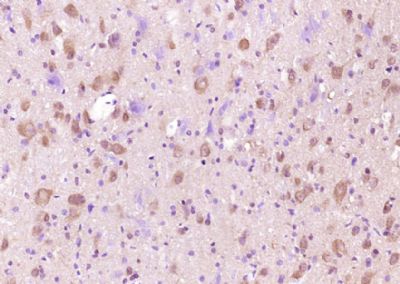
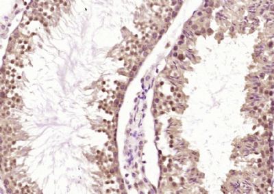
WDFY3 Polyclonal Antibody
Purified Rabbit Polyclonal Antibody (Pab)
- SPECIFICATION
- CITATIONS
- PROTOCOLS
- BACKGROUND

Application
| IHC-P, IHC-F, IF, E |
|---|---|
| Primary Accession | Q8IZQ1 |
| Reactivity | Rat, Pig, Dog, Bovine |
| Host | Rabbit |
| Clonality | Polyclonal |
| Calculated MW | 395 KDa |
| Physical State | Liquid |
| Immunogen | KLH conjugated synthetic peptide derived from human WDFY3 |
| Epitope Specificity | 951-1050/3526 |
| Isotype | IgG |
| Purity | affinity purified by Protein A |
| Buffer | 0.01M TBS (pH7.4) with 1% BSA, 0.02% Proclin300 and 50% Glycerol. |
| SUBCELLULAR LOCATION | Membrane; Peripheral membrane protein; Cytoplasmic side. Note: Colocalizes with autophagic structures in starved cells. |
| SIMILARITY | Contains 1 BEACH domain. Contains 1 FYVE-type zinc finger. Contains 5 WD repeats. |
| Important Note | This product as supplied is intended for research use only, not for use in human, therapeutic or diagnostic applications. |
| Background Descriptions | WDFY3 was identified in a screen for phosphatidylinositol 3 phosphate binding proteins. The FYVE domain is a zinc binding domain found in many PtdIns binding proteins and is proposed to target proteins to membrane lipids. WDFY3 has been shown localized to the nuclear membrane and within autophagic membranes and it is proposed to function as a link between protein aggregates and the autophagic machinery. WDFY3 is also known as autophagy linked FYVE protein. |
| Gene ID | 23001 |
|---|---|
| Other Names | WD repeat and FYVE domain-containing protein 3, Autophagy-linked FYVE protein, Alfy, WDFY3, KIAA0993 |
| Target/Specificity | Ubiquitous. |
| Dilution | IHC-P=1:100-500,IHC-F=1:100-500,IF=1:50-200,ELISA=1:5000-10000 |
| Format | 0.01M TBS(pH7.4), 0.09% (W/V) sodium azide and 50% Glyce |
| Storage | Store at -20 ℃ for one year. Avoid repeated freeze/thaw cycles. When reconstituted in sterile pH 7.4 0.01M PBS or diluent of antibody the antibody is stable for at least two weeks at 2-4 ℃. |
| Name | WDFY3 |
|---|---|
| Synonyms | KIAA0993 |
| Function | Required for selective macroautophagy (aggrephagy). Acts as an adapter protein by linking specific proteins destined for degradation to the core autophagic machinery members, such as the ATG5- ATG12-ATG16L E3-like ligase, SQSTM1 and LC3 (PubMed:20417604). Along with p62/SQSTM1, involved in the formation and autophagic degradation of cytoplasmic ubiquitin-containing inclusions (p62 bodies, ALIS/aggresome-like induced structures). Along with SQSTM1, required to recruit ubiquitinated proteins to PML bodies in the nucleus (PubMed:20168092). Important for normal brain development. Essential for the formation of axonal tracts throughout the brain and spinal cord, including the formation of the major forebrain commissures. Involved in the ability of neural cells to respond to guidance cues. Required for cortical neurons to respond to the trophic effects of netrin-1/NTN1 (By similarity). Regulates Wnt signaling through the removal of DVL3 aggregates, likely in an autophagy-dependent manner. This process may be important for the determination of brain size during embryonic development (PubMed:27008544). May regulate osteoclastogenesis by acting on the TNFSF11/RANKL - TRAF6 pathway (By similarity). After cytokinetic abscission, involved in midbody remnant degradation (PubMed:24128730). In vitro strongly binds to phosphatidylinositol 3-phosphate (PtdIns3P) (PubMed:15292400). |
| Cellular Location | Nucleus membrane. Cytoplasm, cytosol. Nucleus, PML body. Membrane; Peripheral membrane protein; Cytoplasmic side Perikaryon {ECO:0000250|UniProtKB:Q6VNB8}. Cell projection, axon {ECO:0000250|UniProtKB:Q6VNB8}. Note=Relocalization from the nucleus to the cytosol is stimulated by cellular stress, such as starvation or proteasomal inhibition. In the cytosol of starved cells, colocalizes with autophagic structures (PubMed:15292400, PubMed:20168092, PubMed:20417604, PubMed:20971078). This redistribution is dependent on p62/SQSTM1 (PubMed:20168092). When nuclear export is blocked by treatment with leptomycin B, accumulates in nuclear bodies, that completely or partially colocalize with promyelocytic leukemia (PML) bodies (PubMed:20168092). Localizes throughout neurons, including within axons. In neurons, enriched in the light membrane fraction along with the synaptosomal membrane protein synaptophysin and the membrane- bound form of LC3/MAP1LC3A/MAP1LC3B, called LC3-II, a classic marker for autophagic vesicles (By similarity). {ECO:0000250|UniProtKB:Q6VNB8, ECO:0000269|PubMed:15292400, ECO:0000269|PubMed:20168092, ECO:0000269|PubMed:20417604, ECO:0000269|PubMed:20971078} |
| Tissue Location | Expressed in osteoclast and their mononuclear precursors (at protein level). |
Research Areas
Citations (0)

Thousands of laboratories across the world have published research that depended on the performance of antibodies from Abcepta to advance their research. Check out links to articles that cite our products in major peer-reviewed journals, organized by research category.
Submit your citation using an Abcepta antibody to
info@abcepta.com, and receive a free "I Love Antibodies" mug.
info@abcepta.com, and receive a free "I Love Antibodies" mug.

Application Protocols
Provided below are standard protocols that you may find useful for product applications.
Abcepta welcomes feedback from its customers.
If you have used an Abcepta product and would like to share how it has performed, please click on the "Submit Review" button and provide the requested information. Our staff will examine and post your review and contact you if needed.
If you have any additional inquiries please email technical services at tech@abcepta.com.
$ 385.00
Cat# AP59425
Ordering Information
United States
AlbaniaAustraliaAustriaBelgiumBosnia & HerzegovinaBrazilBulgariaCanadaCentral AmericaChinaCroatiaCyprusCzech RepublicDenmarkEstoniaFinlandFranceGermanyGreeceHong KongHungaryIcelandIndiaIndonesiaIrelandIsraelItalyJapanLatviaLithuaniaLuxembourgMacedoniaMalaysiaMaltaMexicoNetherlandsNew ZealandNorwayPakistanPolandPortugalRomaniaSerbiaSingaporeSlovakiaSloveniaSouth AfricaSouth KoreaSpainSwedenSwitzerlandTaiwanTurkeyUnited KingdomUnited StatesVietnamWorldwideOthers
USA Headquarters
(888) 735-7227 / (858) 622-0099 or (858) 875-1900
Other Products
Shipping Information
Domestic orders (in stock items)
Shipped out the same day. Orders placed after 1 PM (PST) will ship out the next business day.
International orders
Contact your local distributors
