


 Foundational characteristics of cancer include proliferation, angiogenesis, migration, evasion of apoptosis, and cellular immortality. Find key markers for these cellular processes and antibodies to detect them.
 The SUMOplot™ Analysis Program predicts and scores sumoylation sites in your protein. SUMOylation is a post-translational modification involved in various cellular processes, such as nuclear-cytosolic transport, transcriptional regulation, apoptosis, protein stability, response to stress, and progression through the cell cycle.
 The Autophagy Receptor Motif Plotter predicts and scores autophagy receptor binding sites in your protein. Identifying proteins connected to this pathway is critical to understanding the role of autophagy in physiological as well as pathological processes such as development, differentiation, neurodegenerative diseases, stress, infection, and cancer.
Anti-JIP3 Antibody
Rabbit polyclonal antibody to JIP3
- SPECIFICATION
- CITATIONS
- PROTOCOLS
- BACKGROUND

Application
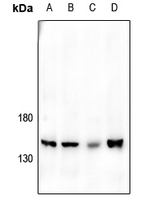
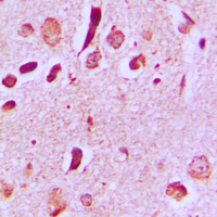
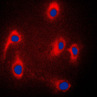
| WB, IF/IC, IHC |
|---|---|
| Primary Accession | Q9UPT6 |
| Other Accession | Q9ESN9 |
| Reactivity | Human, Mouse, Rat, Bovine |
| Host | Rabbit |
| Clonality | Polyclonal |
| Calculated MW | 147457 Da |
| Gene ID | 23162 |
|---|---|
| Other Names | JIP3; KIAA1066; C-Jun-amino-terminal kinase-interacting protein 3; JIP-3; JNK-interacting protein 3; JNK MAP kinase scaffold protein 3; Mitogen-activated protein kinase 8-interacting protein 3 |
| Target/Specificity | KLH-conjugated synthetic peptide encompassing a sequence within the center region of human JIP3. The exact sequence is proprietary. |
| Dilution | WB~~WB (1/500 - 1/1000), IH (1/100 - 1/200), IF/IC (1/100 - 1/500) IF/IC~~N/A IHC~~1:100~500 |
| Format | Liquid in 0.42% Potassium phosphate, 0.87% Sodium chloride, pH 7.3, 30% glycerol, and 0.09% (W/V) sodium azide. |
| Storage | Store at -20 °C.Stable for 12 months from date of receipt |
| Name | MAPK8IP3 |
|---|---|
| Synonyms | JIP3, KIAA1066 |
| Function | The JNK-interacting protein (JIP) group of scaffold proteins selectively mediates JNK signaling by aggregating specific components of the MAPK cascade to form a functional JNK signaling module (PubMed:12189133). May function as a regulator of vesicle transport, through interactions with the JNK-signaling components and motor proteins (By similarity). Promotes neuronal axon elongation in a kinesin- and JNK-dependent manner. Activates cofilin at axon tips via local activation of JNK, thereby regulating filopodial dynamics and enhancing axon elongation. Its binding to kinesin heavy chains (KHC), promotes kinesin-1 motility along microtubules and is essential for axon elongation and regeneration. Regulates cortical neuronal migration by mediating NTRK2/TRKB anterograde axonal transport during brain development (By similarity). Acts as an adapter that bridges the interaction between NTRK2/TRKB and KLC1 and drives NTRK2/TRKB axonal but not dendritic anterograde transport, which is essential for subsequent BDNF-triggered signaling and filopodia formation (PubMed:21775604). |
| Cellular Location | Cytoplasm {ECO:0000250|UniProtKB:Q9ESN9}. Golgi apparatus {ECO:0000250|UniProtKB:Q9ESN9}. Cytoplasmic vesicle {ECO:0000250|UniProtKB:Q9ESN9}. Cell projection, growth cone {ECO:0000250|UniProtKB:Q9ESN9}. Cell projection, axon {ECO:0000250|UniProtKB:E9PSK7}. Cell projection, dendrite {ECO:0000250|UniProtKB:E9PSK7}. Cytoplasm, perinuclear region {ECO:0000250|UniProtKB:E9PSK7}. Note=Localized in the soma and growth cones of differentiated neurites and the Golgi and vesicles of the early secretory compartment of epithelial cells. KIF5A/B/C-mediated transportation to axon tips is essential for its function in enhancing neuronal axon elongation. {ECO:0000250|UniProtKB:E9PSK7, ECO:0000250|UniProtKB:Q9ESN9} |

Thousands of laboratories across the world have published research that depended on the performance of antibodies from Abcepta to advance their research. Check out links to articles that cite our products in major peer-reviewed journals, organized by research category.
info@abcepta.com, and receive a free "I Love Antibodies" mug.

Provided below are standard protocols that you may find useful for product applications.
Background
KLH-conjugated synthetic peptide encompassing a sequence within the center region of human JIP3. The exact sequence is proprietary.
If you have used an Abcepta product and would like to share how it has performed, please click on the "Submit Review" button and provide the requested information. Our staff will examine and post your review and contact you if needed.
If you have any additional inquiries please email technical services at tech@abcepta.com.
Ordering Information
Other Products
Shipping Information
