


 Foundational characteristics of cancer include proliferation, angiogenesis, migration, evasion of apoptosis, and cellular immortality. Find key markers for these cellular processes and antibodies to detect them.
 The SUMOplot™ Analysis Program predicts and scores sumoylation sites in your protein. SUMOylation is a post-translational modification involved in various cellular processes, such as nuclear-cytosolic transport, transcriptional regulation, apoptosis, protein stability, response to stress, and progression through the cell cycle.
 The Autophagy Receptor Motif Plotter predicts and scores autophagy receptor binding sites in your protein. Identifying proteins connected to this pathway is critical to understanding the role of autophagy in physiological as well as pathological processes such as development, differentiation, neurodegenerative diseases, stress, infection, and cancer.
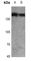
Anti-PHLPP Antibody
Rabbit polyclonal antibody to PHLPP
- SPECIFICATION
- CITATIONS
- PROTOCOLS
- BACKGROUND

Application
| WB |
|---|---|
| Primary Accession | O60346 |
| Other Accession | Q8CHE4 |
| Reactivity | Human, Mouse, Rat |
| Host | Rabbit |
| Clonality | Polyclonal |
| Calculated MW | 184672 Da |
| Gene ID | 23239 |
|---|---|
| Other Names | KIAA0606; PHLPP; PLEKHE1; SCOP; PH domain leucine-rich repeat-containing protein phosphatase 1; Pleckstrin homology domain-containing family E member 1; PH domain-containing family E member 1; Suprachiasmatic nucleus circadian oscillatory protein; hSCOP |
| Target/Specificity | KLH-conjugated synthetic peptide encompassing a sequence within the C-term region of human PHLPP. The exact sequence is proprietary. |
| Dilution | WB~~WB (1/500 - 1/1000) |
| Format | Liquid in 0.42% Potassium phosphate, 0.87% Sodium chloride, pH 7.3, 30% glycerol, and 0.09% (W/V) sodium azide. |
| Storage | Store at -20 °C.Stable for 12 months from date of receipt |
| Name | PHLPP1 |
|---|---|
| Synonyms | KIAA0606, PHLPP, PLEKHE1, SCOP |
| Function | Protein phosphatase involved in regulation of Akt and PKC signaling. Mediates dephosphorylation in the C-terminal domain hydrophobic motif of members of the AGC Ser/Thr protein kinase family; specifically acts on 'Ser-473' of AKT2 and AKT3, 'Ser-660' of PRKCB and 'Ser-657' of PRKCA (PubMed:15808505, PubMed:17386267, PubMed:18162466). Isoform 2 seems to have a major role in regulating Akt signaling in hippocampal neurons (By similarity). Akt regulates the balance between cell survival and apoptosis through a cascade that primarily alters the function of transcription factors that regulate pro- and antiapoptotic genes. Dephosphorylation of 'Ser-473' of Akt triggers apoptosis and suppression of tumor growth. Dephosphorylation of PRKCA and PRKCB leads to their destabilization and degradation (PubMed:18162466). Dephosphorylates STK4 on 'Thr-387' leading to STK4 activation and apoptosis (PubMed:20513427). Dephosphorylates RPS6KB1 and is involved in regulation of cap-dependent translation (PubMed:21986499). Inhibits cancer cell proliferation and may act as a tumor suppressor (PubMed:19079341). Dephosphorylates RAF1 inhibiting its kinase activity (PubMed:24530606). May act as a negative regulator of K-Ras signaling in membrane rafts (By similarity). Involved in the hippocampus- dependent long-term memory formation (By similarity). Involved in circadian control by regulating the consolidation of circadian periodicity after resetting (By similarity). Involved in development and function of regulatory T-cells (By similarity). |
| Cellular Location | Cytoplasm. Membrane; Peripheral membrane protein. Nucleus. Note=In colorectal cancer tissue, expression is concentrated at the lateral membrane of epithelial cells |
| Tissue Location | In colorectal cancer tissue, expression is highest in the surface epithelium of normal colonic mucosa adjacent to the cancer tissue but is largely excluded from the crypt bases. Expression is lost or significantly decreased in 78% of tested tumors (at protein level). Ubiquitously expressed in non-cancerous tissues |

Thousands of laboratories across the world have published research that depended on the performance of antibodies from Abcepta to advance their research. Check out links to articles that cite our products in major peer-reviewed journals, organized by research category.
info@abcepta.com, and receive a free "I Love Antibodies" mug.

Provided below are standard protocols that you may find useful for product applications.
Background
KLH-conjugated synthetic peptide encompassing a sequence within the C-term region of human PHLPP. The exact sequence is proprietary.
If you have used an Abcepta product and would like to share how it has performed, please click on the "Submit Review" button and provide the requested information. Our staff will examine and post your review and contact you if needed.
If you have any additional inquiries please email technical services at tech@abcepta.com.
Ordering Information
Other Products
Shipping Information
