


 Foundational characteristics of cancer include proliferation, angiogenesis, migration, evasion of apoptosis, and cellular immortality. Find key markers for these cellular processes and antibodies to detect them.
 The SUMOplot™ Analysis Program predicts and scores sumoylation sites in your protein. SUMOylation is a post-translational modification involved in various cellular processes, such as nuclear-cytosolic transport, transcriptional regulation, apoptosis, protein stability, response to stress, and progression through the cell cycle.
 The Autophagy Receptor Motif Plotter predicts and scores autophagy receptor binding sites in your protein. Identifying proteins connected to this pathway is critical to understanding the role of autophagy in physiological as well as pathological processes such as development, differentiation, neurodegenerative diseases, stress, infection, and cancer.
Anti-Merlin (pS518) Antibody
Rabbit polyclonal antibody to Merlin (pS518)
- SPECIFICATION
- CITATIONS
- PROTOCOLS
- BACKGROUND

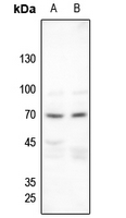
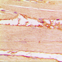
Application
| WB, IHC |
|---|---|
| Primary Accession | P35240 |
| Other Accession | P46662 |
| Reactivity | Human, Mouse, Rat, Chicken |
| Host | Rabbit |
| Clonality | Polyclonal |
| Calculated MW | 69690 Da |
| Gene ID | 4771 |
|---|---|
| Other Names | SCH; Merlin; Moesin-ezrin-radixin-like protein; Neurofibromin-2; Schwannomerlin; Schwannomin |
| Target/Specificity | KLH-conjugated synthetic peptide encompassing a sequence within the C-term region of human Merlin (pS518). The exact sequence is proprietary. |
| Dilution | WB~~WB (1/500 - 1/1000), IH (1/100 - 1/200) IHC~~1:100~500 |
| Format | Liquid in 0.42% Potassium phosphate, 0.87% Sodium chloride, pH 7.3, 30% glycerol, and 0.09% (W/V) sodium azide. |
| Storage | Store at -20 °C.Stable for 12 months from date of receipt |
| Name | NF2 |
|---|---|
| Synonyms | SCH |
| Function | Probable regulator of the Hippo/SWH (Sav/Wts/Hpo) signaling pathway, a signaling pathway that plays a pivotal role in tumor suppression by restricting proliferation and promoting apoptosis. Along with WWC1 can synergistically induce the phosphorylation of LATS1 and LATS2 and can probably function in the regulation of the Hippo/SWH (Sav/Wts/Hpo) signaling pathway. May act as a membrane stabilizing protein. May inhibit PI3 kinase by binding to AGAP2 and impairing its stimulating activity. Suppresses cell proliferation and tumorigenesis by inhibiting the CUL4A-RBX1-DDB1-VprBP/DCAF1 E3 ubiquitin-protein ligase complex. |
| Cellular Location | [Isoform 1]: Cell projection, filopodium membrane; Peripheral membrane protein; Cytoplasmic side. Cell projection, ruffle membrane; Peripheral membrane protein; Cytoplasmic side. Nucleus. Note=In a fibroblastic cell line, isoform 1 is found homogeneously distributed over the entire cell, with a particularly strong staining in ruffling membranes and filopodia. Colocalizes with MPP1 in non-myelin-forming Schwann cells. Binds with DCAF1 in the nucleus. The intramolecular association of the FERM domain with the C- terminal tail promotes nuclear accumulation. The unphosphorylated form accumulates predominantly in the nucleus while the phosphorylated form is largely confined to the non-nuclear fractions [Isoform 9]: Cytoplasm, perinuclear region. Cytoplasmic granule. Note=Observed in cytoplasmic granules concentrated in a perinuclear location. Isoform 9 is absent from ruffling membranes and filopodia |
| Tissue Location | Widely expressed. Isoform 1 and isoform 3 are predominant. Isoform 4, isoform 5 and isoform 6 are expressed moderately. Isoform 8 is found at low frequency. Isoform 7, isoform 9 and isoform 10 are not expressed in adult tissues, with the exception of adult retina expressing isoform 10. Isoform 9 is faintly expressed in fetal brain, heart, lung, skeletal muscle and spleen. Fetal thymus expresses isoforms 1, 7, 9 and 10 at similar levels |

Thousands of laboratories across the world have published research that depended on the performance of antibodies from Abcepta to advance their research. Check out links to articles that cite our products in major peer-reviewed journals, organized by research category.
info@abcepta.com, and receive a free "I Love Antibodies" mug.

Provided below are standard protocols that you may find useful for product applications.
Background
KLH-conjugated synthetic peptide encompassing a sequence within the C-term region of human Merlin (pS518). The exact sequence is proprietary.
If you have used an Abcepta product and would like to share how it has performed, please click on the "Submit Review" button and provide the requested information. Our staff will examine and post your review and contact you if needed.
If you have any additional inquiries please email technical services at tech@abcepta.com.
Ordering Information
Other Products
Shipping Information
