


 Foundational characteristics of cancer include proliferation, angiogenesis, migration, evasion of apoptosis, and cellular immortality. Find key markers for these cellular processes and antibodies to detect them.
 The SUMOplot™ Analysis Program predicts and scores sumoylation sites in your protein. SUMOylation is a post-translational modification involved in various cellular processes, such as nuclear-cytosolic transport, transcriptional regulation, apoptosis, protein stability, response to stress, and progression through the cell cycle.
 The Autophagy Receptor Motif Plotter predicts and scores autophagy receptor binding sites in your protein. Identifying proteins connected to this pathway is critical to understanding the role of autophagy in physiological as well as pathological processes such as development, differentiation, neurodegenerative diseases, stress, infection, and cancer.
Anti-OPA1 Antibody
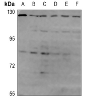
Rabbit polyclonal antibody to OPA1
- SPECIFICATION
- CITATIONS
- PROTOCOLS
- BACKGROUND

Application
| WB |
|---|---|
| Primary Accession | O60313 |
| Other Accession | P58281 |
| Reactivity | Human, Mouse, Rat |
| Host | Rabbit |
| Clonality | Polyclonal |
| Calculated MW | 111631 Da |
| Gene ID | 4976 |
|---|---|
| Other Names | KIAA0567; Dynamin-like 120 kDa protein mitochondrial; Optic atrophy protein 1 |
| Target/Specificity | KLH-conjugated synthetic peptide encompassing a sequence within the C-term region of human OPA1. The exact sequence is proprietary. |
| Dilution | WB~~WB (1/500 - 1/1000) |
| Format | Liquid in 0.42% Potassium phosphate, 0.87% Sodium chloride, pH 7.3, 30% glycerol, and 0.09% (W/V) sodium azide. |
| Storage | Store at -20 °C.Stable for 12 months from date of receipt |
| Name | OPA1 |
|---|---|
| Function | Dynamin-related GTPase that is essential for normal mitochondrial morphology by mediating fusion of the mitochondrial inner membranes, regulating cristae morphology and maintaining respiratory chain function (PubMed:16778770, PubMed:17709429, PubMed:20185555, PubMed:24616225, PubMed:28628083, PubMed:28746876, PubMed:31922487, PubMed:32228866, PubMed:32567732, PubMed:33130824, PubMed:33237841, PubMed:37612504, PubMed:37612506). Exists in two forms: the transmembrane, long form (Dynamin-like GTPase OPA1, long form; L-OPA1), which is tethered to the inner mitochondrial membrane, and the short soluble form (Dynamin-like GTPase OPA1, short form; S-OPA1), which results from proteolytic cleavage and localizes in the intermembrane space (PubMed:31922487, PubMed:32228866, PubMed:33237841, PubMed:37612504, PubMed:37612506). Both forms (L-OPA1 and S-OPA1) cooperate to catalyze the fusion of the mitochondrial inner membrane (PubMed:31922487, PubMed:37612504, PubMed:37612506). The equilibrium between L-OPA1 and S-OPA1 is essential: excess levels of S-OPA1, produced by cleavage by OMA1 following loss of mitochondrial membrane potential, lead to an impaired equilibrium between L-OPA1 and S-OPA1, inhibiting mitochondrial fusion (PubMed:20038677, PubMed:31922487). The balance between L-OPA1 and S-OPA1 also influences cristae shape and morphology (By similarity). Involved in remodeling cristae and the release of cytochrome c during apoptosis (By similarity). Proteolytic processing by PARL in response to intrinsic apoptotic signals may lead to disassembly of OPA1 oligomers and release of the caspase activator cytochrome C (CYCS) into the mitochondrial intermembrane space (By similarity). Acts as a regulator of T-helper Th17 cells, which are characterized by cells with fused mitochondria with tight cristae, by mediating mitochondrial membrane remodeling: OPA1 is required for interleukin-17 (IL-17) production (By similarity). Its role in mitochondrial morphology is required for mitochondrial genome maintenance (PubMed:18158317, PubMed:20974897). |
| Cellular Location | [Dynamin-like GTPase OPA1, long form]: Mitochondrion inner membrane; Single-pass membrane protein. Note=Detected at contact sites between endoplasmic reticulum and mitochondrion membranes. |
| Tissue Location | Highly expressed in retina (PubMed:11017079, PubMed:11017080, PubMed:11810270). Also expressed in brain, testis, heart and skeletal muscle (PubMed:11810270). Low levels of all isoforms expressed in a variety of tissues (PubMed:11810270) [Isoform 2]: Isoform 2 expressed in colon, liver, kidney, thyroid gland and leukocytes. |

Thousands of laboratories across the world have published research that depended on the performance of antibodies from Abcepta to advance their research. Check out links to articles that cite our products in major peer-reviewed journals, organized by research category.
info@abcepta.com, and receive a free "I Love Antibodies" mug.

Provided below are standard protocols that you may find useful for product applications.
Background
KLH-conjugated synthetic peptide encompassing a sequence within the C-term region of human OPA1. The exact sequence is proprietary.
If you have used an Abcepta product and would like to share how it has performed, please click on the "Submit Review" button and provide the requested information. Our staff will examine and post your review and contact you if needed.
If you have any additional inquiries please email technical services at tech@abcepta.com.
Ordering Information
Other Products
Shipping Information
