


 Foundational characteristics of cancer include proliferation, angiogenesis, migration, evasion of apoptosis, and cellular immortality. Find key markers for these cellular processes and antibodies to detect them.
 The SUMOplot™ Analysis Program predicts and scores sumoylation sites in your protein. SUMOylation is a post-translational modification involved in various cellular processes, such as nuclear-cytosolic transport, transcriptional regulation, apoptosis, protein stability, response to stress, and progression through the cell cycle.
 The Autophagy Receptor Motif Plotter predicts and scores autophagy receptor binding sites in your protein. Identifying proteins connected to this pathway is critical to understanding the role of autophagy in physiological as well as pathological processes such as development, differentiation, neurodegenerative diseases, stress, infection, and cancer.
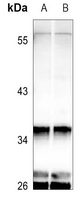
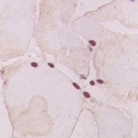
Anti-GNAT1 Antibody
Rabbit polyclonal antibody to GNAT1
- SPECIFICATION
- CITATIONS
- PROTOCOLS
- BACKGROUND

Application
| WB, IHC |
|---|---|
| Primary Accession | P11488 |
| Other Accession | P20612 |
| Reactivity | Human, Mouse, Rat, Bovine, Dog |
| Host | Rabbit |
| Clonality | Polyclonal |
| Calculated MW | 40041 Da |
| Gene ID | 2779 |
|---|---|
| Other Names | GNATR; Guanine nucleotide-binding protein G(t) subunit alpha-1; Transducin alpha-1 chain |
| Target/Specificity | KLH-conjugated synthetic peptide encompassing a sequence within the center region of human GNAT1. The exact sequence is proprietary. |
| Dilution | WB~~WB (1/500 - 1/1000), IH (1/100 - 1/200) IHC~~1:100~500 |
| Format | Liquid in 0.42% Potassium phosphate, 0.87% Sodium chloride, pH 7.3, 30% glycerol, and 0.09% (W/V) sodium azide. |
| Storage | Store at -20 °C.Stable for 12 months from date of receipt |
| Name | GNAT1 |
|---|---|
| Synonyms | GNATR |
| Function | Functions as a signal transducer for the rod photoreceptor RHO. Required for normal RHO-mediated light perception by the retina (PubMed:22190596). Guanine nucleotide-binding proteins (G proteins) function as transducers downstream of G protein-coupled receptors (GPCRs), such as the photoreceptor RHO. The alpha chain contains the guanine nucleotide binding site and alternates between an active, GTP- bound state and an inactive, GDP-bound state. Activated RHO promotes GDP release and GTP binding. Signaling is mediated via downstream effector proteins, such as cGMP-phosphodiesterase (By similarity). |
| Cellular Location | Cell projection, cilium, photoreceptor outer segment {ECO:0000250|UniProtKB:P04695}. Membrane {ECO:0000250|UniProtKB:P04695}; Peripheral membrane protein {ECO:0000250|UniProtKB:P04695}. Photoreceptor inner segment {ECO:0000250|UniProtKB:P20612}. Note=Localizes mainly in the outer segment in the dark-adapted state, whereas is translocated to the inner part of the photoreceptors in the light-adapted state. During dark- adapted conditions, in the presence of UNC119 mislocalizes from the outer segment to the inner part of rod photoreceptors which leads to decreased photoreceptor damage caused by light {ECO:0000250|UniProtKB:P20612} |
| Tissue Location | Rod photoreceptor cells (PubMed:1614872). Predominantly expressed in the retina followed by the ciliary body, iris and retinal pigment epithelium (PubMed:22190596) |

Thousands of laboratories across the world have published research that depended on the performance of antibodies from Abcepta to advance their research. Check out links to articles that cite our products in major peer-reviewed journals, organized by research category.
info@abcepta.com, and receive a free "I Love Antibodies" mug.

Provided below are standard protocols that you may find useful for product applications.
Background
KLH-conjugated synthetic peptide encompassing a sequence within the center region of human GNAT1. The exact sequence is proprietary.
If you have used an Abcepta product and would like to share how it has performed, please click on the "Submit Review" button and provide the requested information. Our staff will examine and post your review and contact you if needed.
If you have any additional inquiries please email technical services at tech@abcepta.com.
Ordering Information
Other Products
Shipping Information
