


 Foundational characteristics of cancer include proliferation, angiogenesis, migration, evasion of apoptosis, and cellular immortality. Find key markers for these cellular processes and antibodies to detect them.
 The SUMOplot™ Analysis Program predicts and scores sumoylation sites in your protein. SUMOylation is a post-translational modification involved in various cellular processes, such as nuclear-cytosolic transport, transcriptional regulation, apoptosis, protein stability, response to stress, and progression through the cell cycle.
 The Autophagy Receptor Motif Plotter predicts and scores autophagy receptor binding sites in your protein. Identifying proteins connected to this pathway is critical to understanding the role of autophagy in physiological as well as pathological processes such as development, differentiation, neurodegenerative diseases, stress, infection, and cancer.
Anti-MAF1 Antibody
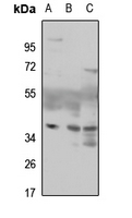
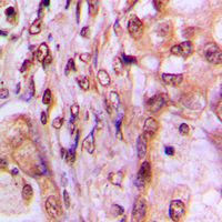
Rabbit polyclonal antibody to MAF1
- SPECIFICATION
- CITATIONS
- PROTOCOLS
- BACKGROUND

Application
| WB, IHC |
|---|---|
| Primary Accession | Q9H063 |
| Other Accession | Q9D0U6 |
| Reactivity | Human, Mouse, Rat, Bovine |
| Host | Rabbit |
| Clonality | Polyclonal |
| Calculated MW | 28771 Da |
| Gene ID | 84232 |
|---|---|
| Other Names | Repressor of RNA polymerase III transcription MAF1 homolog |
| Target/Specificity | KLH-conjugated synthetic peptide encompassing a sequence within the center region of human MAF1. The exact sequence is proprietary. |
| Dilution | WB~~WB (1/500 - 1/1000), IH (1/100 - 1/200) IHC~~1:100~500 |
| Format | Liquid in 0.42% Potassium phosphate, 0.87% Sodium chloride, pH 7.3, 30% glycerol, and 0.09% (W/V) sodium azide. |
| Storage | Store at -20 °C.Stable for 12 months from date of receipt |
| Name | MAF1 |
|---|---|
| Function | Plays a role in the repression of RNA polymerase III-mediated transcription in response to changing nutritional, environmental and cellular stress conditions to balance the production of highly abundant tRNAs, 5S rRNA, and other small non-coding RNAs with cell growth and maintenance (PubMed:18377933, PubMed:20233713, PubMed:20516213, PubMed:20543138). Also plays a key role in cell fate determination by promoting mesorderm induction and adipocyte differentiation (By similarity). Mechanistically, associates with the RNA polymerase III clamp and thereby impairs its recruitment to the complex made of the promoter DNA, TBP and the initiation factor TFIIIB (PubMed:17505538, PubMed:20887893). When nutrients are available and mTOR kinase is active, MAF1 is hyperphosphorylated and RNA polymerase III is engaged in transcription. Stress-induced MAF1 dephosphorylation results in nuclear localization, increased targeting of gene-bound RNA polymerase III and a decrease in the transcriptional readout (PubMed:26941251). Additionally, may also regulate RNA polymerase I and RNA polymerase II- dependent transcription through its ability to regulate expression of the central initiation factor TBP (PubMed:17499043). |
| Cellular Location | Nucleus. Cytoplasm |

Thousands of laboratories across the world have published research that depended on the performance of antibodies from Abcepta to advance their research. Check out links to articles that cite our products in major peer-reviewed journals, organized by research category.
info@abcepta.com, and receive a free "I Love Antibodies" mug.

Provided below are standard protocols that you may find useful for product applications.
Background
KLH-conjugated synthetic peptide encompassing a sequence within the center region of human MAF1. The exact sequence is proprietary.
If you have used an Abcepta product and would like to share how it has performed, please click on the "Submit Review" button and provide the requested information. Our staff will examine and post your review and contact you if needed.
If you have any additional inquiries please email technical services at tech@abcepta.com.
Ordering Information
Other Products
Shipping Information
