


 Foundational characteristics of cancer include proliferation, angiogenesis, migration, evasion of apoptosis, and cellular immortality. Find key markers for these cellular processes and antibodies to detect them.
 The SUMOplot™ Analysis Program predicts and scores sumoylation sites in your protein. SUMOylation is a post-translational modification involved in various cellular processes, such as nuclear-cytosolic transport, transcriptional regulation, apoptosis, protein stability, response to stress, and progression through the cell cycle.
 The Autophagy Receptor Motif Plotter predicts and scores autophagy receptor binding sites in your protein. Identifying proteins connected to this pathway is critical to understanding the role of autophagy in physiological as well as pathological processes such as development, differentiation, neurodegenerative diseases, stress, infection, and cancer.
Anti-MARCH1 Antibody
Rabbit polyclonal antibody to MARCH1
- SPECIFICATION
- CITATIONS
- PROTOCOLS
- BACKGROUND

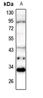
Application
| WB, IHC |
|---|---|
| Primary Accession | Q8TCQ1 |
| Other Accession | Q6NZQ8 |
| Reactivity | Human, Mouse |
| Host | Rabbit |
| Clonality | Polyclonal |
| Calculated MW | 32308 Da |
| Gene ID | 55016 |
|---|---|
| Other Names | RNF171; E3 ubiquitin-protein ligase MARCH1; Membrane-associated RING finger protein 1; Membrane-associated RING-CH protein I; MARCH-I; RING finger protein 171 |
| Target/Specificity | KLH-conjugated synthetic peptide encompassing a sequence within the center region of human MARCH1. The exact sequence is proprietary. |
| Dilution | WB~~WB (1/500 - 1/1000), IH (1/100 - 1/200) IHC~~1:100~500 |
| Format | Liquid in 0.42% Potassium phosphate, 0.87% Sodium chloride, pH 7.3, 30% glycerol, and 0.09% (W/V) sodium azide. |
| Storage | Store at -20 °C.Stable for 12 months from date of receipt |
| Name | MARCHF1 (HGNC:26077) |
|---|---|
| Synonyms | MARCH1, RNF171 |
| Function | E3 ubiquitin-protein ligase that mediates ubiquitination of TFRC, CD86, FAS and MHC class II proteins, such as HLA-DR alpha and beta, and promotes their subsequent endocytosis and sorting to lysosomes via multivesicular bodies (PubMed:18389477, PubMed:18305173, PubMed:21220452, PubMed:35045264). By constitutively ubiquitinating MHC class II proteins in immature dendritic cells, down-regulates their cell surface localization thus sequestering them in the intracellular endosomal system. Also regulates insulin sensitivity by controlling surface expression of the insulin receptor subunit beta/INSR by direct ubiquitination and degradation (PubMed:27577745). |
| Cellular Location | Golgi apparatus, trans-Golgi network membrane {ECO:0000250|UniProtKB:Q6NZQ8}; Multi-pass membrane protein. Lysosome membrane; Multi- pass membrane protein. Cytoplasmic vesicle membrane; Multi-pass membrane protein. Late endosome membrane; Multi-pass membrane protein. Early endosome membrane; Multi-pass membrane protein. Cell membrane; Multi-pass membrane protein |
| Tissue Location | Expressed in antigen presenting cells, APCs, located in lymph nodes and spleen. Also expressed in lung. Expression is high in follicular B-cells, moderate in dendritic cells and low in splenic T-cells. |

Thousands of laboratories across the world have published research that depended on the performance of antibodies from Abcepta to advance their research. Check out links to articles that cite our products in major peer-reviewed journals, organized by research category.
info@abcepta.com, and receive a free "I Love Antibodies" mug.

Provided below are standard protocols that you may find useful for product applications.
Background
KLH-conjugated synthetic peptide encompassing a sequence within the center region of human MARCH1. The exact sequence is proprietary.
If you have used an Abcepta product and would like to share how it has performed, please click on the "Submit Review" button and provide the requested information. Our staff will examine and post your review and contact you if needed.
If you have any additional inquiries please email technical services at tech@abcepta.com.
Ordering Information
Other Products
Shipping Information
