


 Foundational characteristics of cancer include proliferation, angiogenesis, migration, evasion of apoptosis, and cellular immortality. Find key markers for these cellular processes and antibodies to detect them.
 The SUMOplot™ Analysis Program predicts and scores sumoylation sites in your protein. SUMOylation is a post-translational modification involved in various cellular processes, such as nuclear-cytosolic transport, transcriptional regulation, apoptosis, protein stability, response to stress, and progression through the cell cycle.
 The Autophagy Receptor Motif Plotter predicts and scores autophagy receptor binding sites in your protein. Identifying proteins connected to this pathway is critical to understanding the role of autophagy in physiological as well as pathological processes such as development, differentiation, neurodegenerative diseases, stress, infection, and cancer.
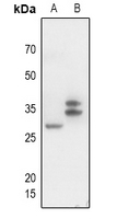
Anti-CD300f Antibody
Rabbit polyclonal antibody to CD300f
- SPECIFICATION
- CITATIONS
- PROTOCOLS
- BACKGROUND

Application
| WB |
|---|---|
| Primary Accession | Q8TDQ1 |
| Reactivity | Human, Mouse, Rat |
| Host | Rabbit |
| Clonality | Polyclonal |
| Calculated MW | 32335 Da |
| Gene ID | 146722 |
|---|---|
| Other Names | CD300F; CLM1; IGSF13; IREM1; NKIR; CMRF35-like molecule 1; CLM-1; CD300 antigen-like family member F; Immune receptor expressed on myeloid cells 1; IREM-1; Immunoglobulin superfamily member 13; IgSF13; NK inhibitory receptor; CD300f |
| Target/Specificity | KLH-conjugated synthetic peptide encompassing a sequence within the center region of human CD300f. The exact sequence is proprietary. |
| Dilution | WB~~WB (1/500 - 1/1000) |
| Format | Liquid in 0.42% Potassium phosphate, 0.87% Sodium chloride, pH 7.3, 30% glycerol, and 0.09% (W/V) sodium azide. |
| Storage | Store at -20 °C.Stable for 12 months from date of receipt |
| Name | CD300LF |
|---|---|
| Synonyms | CD300F, CLM1, IGSF13, IREM1, NKIR |
| Function | Acts as an inhibitory receptor for myeloid cells and mast cells (PubMed:15549731). Positively regulates the phagocytosis of apoptotic cells (efferocytosis) via phosphatidylserine (PS) recognition; recognizes and binds PS as a ligand which is expressed on the surface of apoptotic cells. Plays an important role in the maintenance of immune homeostasis, by promoting macrophage-mediated efferocytosis and by inhibiting dendritic cell-mediated efferocytosis (By similarity). Negatively regulates Fc epsilon receptor-dependent mast cell activation and allergic responses via binding to ceramide and sphingomyelin which act as ligands (PubMed:24035150). May act as a coreceptor for interleukin 4 (IL-4). Associates with and regulates IL-4 receptor alpha-mediated responses by augmenting IL-4- and IL-13-induced signaling (By similarity). Negatively regulates the Toll-like receptor (TLR) signaling mediated by MYD88 and TRIF through activation of PTPN6/SHP-1 and PTPN11/SHP-2 (PubMed:22043923). Inhibits osteoclast formation. Induces macrophage cell death upon engagement (By similarity). |
| Cellular Location | Cell membrane; Single-pass type I membrane protein |
| Tissue Location | Highly expressed in spleen, peripheral blood leukocyte and monocyte, and lung. Weakly expressed in thymus, heart, brain, placenta, liver, skeletal muscle, kidney, pancreas, prostate, testis, ovary, small intestine or colon. Expressed selectively in monocytes and monocyte-related cells. |

Thousands of laboratories across the world have published research that depended on the performance of antibodies from Abcepta to advance their research. Check out links to articles that cite our products in major peer-reviewed journals, organized by research category.
info@abcepta.com, and receive a free "I Love Antibodies" mug.

Provided below are standard protocols that you may find useful for product applications.
Background
KLH-conjugated synthetic peptide encompassing a sequence within the center region of human CD300f. The exact sequence is proprietary.
If you have used an Abcepta product and would like to share how it has performed, please click on the "Submit Review" button and provide the requested information. Our staff will examine and post your review and contact you if needed.
If you have any additional inquiries please email technical services at tech@abcepta.com.
Ordering Information
Other Products
Shipping Information
