


 Foundational characteristics of cancer include proliferation, angiogenesis, migration, evasion of apoptosis, and cellular immortality. Find key markers for these cellular processes and antibodies to detect them.
 The SUMOplot™ Analysis Program predicts and scores sumoylation sites in your protein. SUMOylation is a post-translational modification involved in various cellular processes, such as nuclear-cytosolic transport, transcriptional regulation, apoptosis, protein stability, response to stress, and progression through the cell cycle.
 The Autophagy Receptor Motif Plotter predicts and scores autophagy receptor binding sites in your protein. Identifying proteins connected to this pathway is critical to understanding the role of autophagy in physiological as well as pathological processes such as development, differentiation, neurodegenerative diseases, stress, infection, and cancer.
Anti-CD316 Antibody
Rabbit polyclonal antibody to CD316
- SPECIFICATION
- CITATIONS
- PROTOCOLS
- BACKGROUND

Application
| WB |
|---|---|
| Primary Accession | Q969P0 |
| Other Accession | Q8R366 |
| Reactivity | Human, Mouse |
| Host | Rabbit |
| Clonality | Polyclonal |
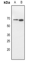
| Calculated MW | 65034 Da |
| Gene ID | 93185 |
|---|---|
| Other Names | CD81P3; EWI2; KCT4; Immunoglobulin superfamily member 8; IgSF8; CD81 partner 3; Glu-Trp-Ile EWI motif-containing protein 2; EWI-2; Keratinocytes-associated transmembrane protein 4; KCT-4; LIR-D1; Prostaglandin regulatory-like protein; PGRL; CD316 |
| Target/Specificity | KLH-conjugated synthetic peptide encompassing a sequence within the center region of human CD316. The exact sequence is proprietary. |
| Dilution | WB~~WB (1/500 - 1/1000) |
| Format | Liquid in 0.42% Potassium phosphate, 0.87% Sodium chloride, pH 7.3, 30% glycerol, and 0.09% (W/V) sodium azide. |
| Storage | Store at -20 °C.Stable for 12 months from date of receipt |
| Name | IGSF8 |
|---|---|
| Synonyms | CD81P3, EWI2, KCT4 |
| Function | Member of the immunoglobulin superfamily (IgSF) that links tetraspanin-enriched microdomains to the actin cytoskeleton and plays several important roles in innate and adaptive immunity (PubMed:11504738, PubMed:14662754). Acts as an inducible receptor of HSPA8 on dendritic cells to enhance the CCL21/SLC-dependent migration of activated mature dendritic cells while attenuating their antigen- specific stimulatory capacities (PubMed:17785435). In complex with alpha-actinins ACTN1 and ACTN4, regulates actin dynamics in the immune synapse and subsequent T-cell activation (PubMed:22689882). Inhibits the entry of several viruses such as hepatitis C Virus (HCV) or HIV-1. Mechanistically, promotes a change in CD81 organization at the plasma membrane by significantly restricting its diffusion which in turn influences CD81 interaction with Claudin-1/CLDN1, preventing CLDN1 from acting as a co-receptor required for HCV entry (PubMed:23351194). Accumulates at the presynaptic terminal, the producer cell side of the virological synapse, to prevent HIV-1 Env-mediated cell-cell fusion (PubMed:31757023). Highly expressed on malignant cells with antigen presentation defects, interacts with NK receptor KIR3DL2 to suppress NK-cell cytotoxicity (PubMed:38657602). May participate in the regulation of neurite outgrowth and maintenance of the neural network in the adult brain. |
| Cellular Location | Cell membrane; Single-pass membrane protein. Note=Colocalizes with CD81 at the immune synapse. |
| Tissue Location | Expressed in brain, kidney, testis, liver and placenta with moderate expression in all other tissues. Detected on a majority of B-cells, T-cells, and natural killer cells (PubMed:12708969). Expressed on dendritic cells (PubMed:17785435) |

Thousands of laboratories across the world have published research that depended on the performance of antibodies from Abcepta to advance their research. Check out links to articles that cite our products in major peer-reviewed journals, organized by research category.
info@abcepta.com, and receive a free "I Love Antibodies" mug.

Provided below are standard protocols that you may find useful for product applications.
Background
KLH-conjugated synthetic peptide encompassing a sequence within the center region of human CD316. The exact sequence is proprietary.
If you have used an Abcepta product and would like to share how it has performed, please click on the "Submit Review" button and provide the requested information. Our staff will examine and post your review and contact you if needed.
If you have any additional inquiries please email technical services at tech@abcepta.com.
Ordering Information
Other Products
Shipping Information
