


 Foundational characteristics of cancer include proliferation, angiogenesis, migration, evasion of apoptosis, and cellular immortality. Find key markers for these cellular processes and antibodies to detect them.
 The SUMOplot™ Analysis Program predicts and scores sumoylation sites in your protein. SUMOylation is a post-translational modification involved in various cellular processes, such as nuclear-cytosolic transport, transcriptional regulation, apoptosis, protein stability, response to stress, and progression through the cell cycle.
 The Autophagy Receptor Motif Plotter predicts and scores autophagy receptor binding sites in your protein. Identifying proteins connected to this pathway is critical to understanding the role of autophagy in physiological as well as pathological processes such as development, differentiation, neurodegenerative diseases, stress, infection, and cancer.
Anti-RGS14 Antibody
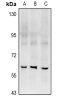
Rabbit polyclonal antibody to RGS14
- SPECIFICATION
- CITATIONS
- PROTOCOLS
- BACKGROUND

Application
| WB |
|---|---|
| Primary Accession | O43566 |
| Other Accession | P97492 |
| Reactivity | Human, Mouse, Rat |
| Host | Rabbit |
| Clonality | Polyclonal |
| Calculated MW | 61447 Da |
| Gene ID | 10636 |
|---|---|
| Other Names | Regulator of G-protein signaling 14; RGS14 |
| Target/Specificity | KLH-conjugated synthetic peptide encompassing a sequence within the center region of human RGS14. The exact sequence is proprietary. |
| Dilution | WB~~WB (1/500 - 1/1000) |
| Format | Liquid in 0.42% Potassium phosphate, 0.87% Sodium chloride, pH 7.3, 30% glycerol, and 0.09% (W/V) sodium azide. |
| Storage | Store at -20 °C.Stable for 12 months from date of receipt |
| Name | RGS14 |
|---|---|
| Function | Regulates G protein-coupled receptor signaling cascades. Inhibits signal transduction by increasing the GTPase activity of G protein alpha subunits, thereby driving them into their inactive GDP- bound form. Besides, modulates signal transduction via G protein alpha subunits by functioning as a GDP-dissociation inhibitor (GDI). Has GDI activity on G(i) alpha subunits GNAI1 and GNAI3, but not on GNAI2 and G(o)-alpha subunit GNAO1. Has GAP activity on GNAI0, GNAI2 and GNAI3. May act as a scaffold integrating G protein and Ras/Raf MAPkinase signaling pathways. Inhibits platelet-derived growth factor (PDGF)- stimulated ERK1/ERK2 phosphorylation; a process depending on its interaction with HRAS and that is reversed by G(i) alpha subunit GNAI1. Acts as a positive modulator of microtubule polymerisation and spindle organization through a G(i)-alpha-dependent mechanism. Plays a role in cell division. Required for the nerve growth factor (NGF)-mediated neurite outgrowth. Involved in stress resistance. May be involved in visual memory processing capacity and hippocampal-based learning and memory. |
| Cellular Location | Nucleus. Nucleus, PML body. Cytoplasm. Membrane. Cell membrane. Cytoplasm, cytoskeleton, microtubule organizing center, centrosome. Cytoplasm, cytoskeleton, spindle. Cytoplasm, cytoskeleton, spindle pole. Cell projection, dendrite. Cell projection, dendritic spine Postsynaptic density. Note=Associates with the perinuclear sheaths of microtubules (MTs) surrounding the pronuclei, prior to segregating to the anastral mitotic apparatus and subsequently the barrel-shaped cytoplasmic bridge between the nascent nuclei of the emerging 2-cell embryo. Localizes to a perinuclear compartment near the microtubule-organizing center (MTOC). Expressed in the nucleus during interphase and segregates to the centrosomes and astral MTs during mitosis. Relocalizes to the nucleus in PML nuclear bodies in response to heat stress. Colocalizes with RIC8A in CA2 hippocampal neurons Localizes to spindle poles during metaphase. Shuttles between the nucleus and cytoplasm in a CRM1-dependent manner. Recruited from the cytosol to the plasma membrane by the inactive GDP-bound forms of G(i) alpha subunits GNAI1 and GNAI3. Recruited from the cytosol to membranes by the active GTP-bound form of HRAS. Colocalizes with G(i) alpha subunit GNAI1 and RIC8A at the plasma membrane. Colocalizes with BRAF and RAF1 in both the cytoplasm and membranes (By similarity) |

Thousands of laboratories across the world have published research that depended on the performance of antibodies from Abcepta to advance their research. Check out links to articles that cite our products in major peer-reviewed journals, organized by research category.
info@abcepta.com, and receive a free "I Love Antibodies" mug.

Provided below are standard protocols that you may find useful for product applications.
Background
KLH-conjugated synthetic peptide encompassing a sequence within the center region of human RGS14. The exact sequence is proprietary.
If you have used an Abcepta product and would like to share how it has performed, please click on the "Submit Review" button and provide the requested information. Our staff will examine and post your review and contact you if needed.
If you have any additional inquiries please email technical services at tech@abcepta.com.
Ordering Information
Other Products
Shipping Information
