


 Foundational characteristics of cancer include proliferation, angiogenesis, migration, evasion of apoptosis, and cellular immortality. Find key markers for these cellular processes and antibodies to detect them.
 The SUMOplot™ Analysis Program predicts and scores sumoylation sites in your protein. SUMOylation is a post-translational modification involved in various cellular processes, such as nuclear-cytosolic transport, transcriptional regulation, apoptosis, protein stability, response to stress, and progression through the cell cycle.
 The Autophagy Receptor Motif Plotter predicts and scores autophagy receptor binding sites in your protein. Identifying proteins connected to this pathway is critical to understanding the role of autophagy in physiological as well as pathological processes such as development, differentiation, neurodegenerative diseases, stress, infection, and cancer.
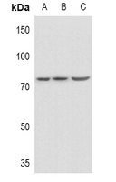
Anti-PLK2 Antibody
Rabbit polyclonal antibody to PLK2
- SPECIFICATION
- CITATIONS
- PROTOCOLS
- BACKGROUND

Application
| WB |
|---|---|
| Primary Accession | Q9NYY3 |
| Reactivity | Human |
| Host | Rabbit |
| Clonality | Polyclonal |
| Calculated MW | 78237 Da |
| Gene ID | 10769 |
|---|---|
| Other Names | SNK; Serine/threonine-protein kinase PLK2; Polo-like kinase 2; PLK-2; hPlk2; Serine/threonine-protein kinase SNK; hSNK; Serum-inducible kinase |
| Target/Specificity | KLH-conjugated synthetic peptide encompassing a sequence within the center region of human PLK2. The exact sequence is proprietary. |
| Dilution | WB~~WB (1/500 - 1/1000) |
| Format | Liquid in 0.42% Potassium phosphate, 0.87% Sodium chloride, pH 7.3, 30% glycerol, and 0.09% (W/V) sodium azide. |
| Storage | Store at -20 °C.Stable for 12 months from date of receipt |
| Name | PLK2 |
|---|---|
| Synonyms | SNK |
| Function | Tumor suppressor serine/threonine-protein kinase involved in synaptic plasticity, centriole duplication and G1/S phase transition. Polo-like kinases act by binding and phosphorylating proteins that are already phosphorylated on a specific motif recognized by the POLO box domains. Phosphorylates CPAP, NPM1, RAPGEF2, RASGRF1, SNCA, SIPA1L1 and SYNGAP1. Plays a key role in synaptic plasticity and memory by regulating the Ras and Rap protein signaling: required for overactivity-dependent spine remodeling by phosphorylating the Ras activator RASGRF1 and the Rap inhibitor SIPA1L1 leading to their degradation by the proteasome. Conversely, phosphorylates the Rap activator RAPGEF2 and the Ras inhibitor SYNGAP1, promoting their activity. Also regulates synaptic plasticity independently of kinase activity, via its interaction with NSF that disrupts the interaction between NSF and the GRIA2 subunit of AMPARs, leading to a rapid rundown of AMPAR-mediated current that occludes long term depression. Required for procentriole formation and centriole duplication by phosphorylating CPAP and NPM1, respectively. Its induction by p53/TP53 suggests that it may participate in the mitotic checkpoint following stress. |
| Cellular Location | Cytoplasm, cytoskeleton, microtubule organizing center, centrosome, centriole. Cell projection, dendrite Note=Localizes to centrosomes during early G1 phase where it only associates to the mother centriole and then distributes equally to both mother and daughter centrioles at the onset of S phase |
| Tissue Location | Expressed at higher level in the fetal lung, kidney, spleen and heart. |

Thousands of laboratories across the world have published research that depended on the performance of antibodies from Abcepta to advance their research. Check out links to articles that cite our products in major peer-reviewed journals, organized by research category.
info@abcepta.com, and receive a free "I Love Antibodies" mug.

Provided below are standard protocols that you may find useful for product applications.
Background
KLH-conjugated synthetic peptide encompassing a sequence within the center region of human PLK2. The exact sequence is proprietary.
If you have used an Abcepta product and would like to share how it has performed, please click on the "Submit Review" button and provide the requested information. Our staff will examine and post your review and contact you if needed.
If you have any additional inquiries please email technical services at tech@abcepta.com.
Ordering Information
Other Products
Shipping Information
