


 Foundational characteristics of cancer include proliferation, angiogenesis, migration, evasion of apoptosis, and cellular immortality. Find key markers for these cellular processes and antibodies to detect them.
 The SUMOplot™ Analysis Program predicts and scores sumoylation sites in your protein. SUMOylation is a post-translational modification involved in various cellular processes, such as nuclear-cytosolic transport, transcriptional regulation, apoptosis, protein stability, response to stress, and progression through the cell cycle.
 The Autophagy Receptor Motif Plotter predicts and scores autophagy receptor binding sites in your protein. Identifying proteins connected to this pathway is critical to understanding the role of autophagy in physiological as well as pathological processes such as development, differentiation, neurodegenerative diseases, stress, infection, and cancer.
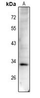
Anti-Cyclin D1 (pT288) Antibody
Rabbit polyclonal antibody to Cyclin D1 (pT288)
- SPECIFICATION
- CITATIONS
- PROTOCOLS
- BACKGROUND

Application
| WB |
|---|---|
| Primary Accession | P24385 |
| Other Accession | P25322 |
| Reactivity | Human, Mouse, Rat |
| Host | Rabbit |
| Clonality | Polyclonal |
| Calculated MW | 33729 Da |
| Gene ID | 595 |
|---|---|
| Other Names | BCL1; PRAD1; G1/S-specific cyclin-D1; B-cell lymphoma 1 protein; BCL-1; BCL-1 oncogene; PRAD1 oncogene |
| Target/Specificity | KLH-conjugated synthetic peptide encompassing a sequence within the C-term region of human Cyclin D1. The exact sequence is proprietary. |
| Dilution | WB~~WB (1/500 - 1/1000) |
| Format | Liquid in 0.42% Potassium phosphate, 0.87% Sodium chloride, pH 7.3, 30% glycerol, and 0.09% (W/V) sodium azide. |
| Storage | Store at -20 °C.Stable for 12 months from date of receipt |
| Name | CCND1 {ECO:0000303|PubMed:8204893, ECO:0000312|HGNC:HGNC:1582} |
|---|---|
| Function | Regulatory component of the cyclin D1-CDK4 (DC) complex that phosphorylates and inhibits members of the retinoblastoma (RB) protein family including RB1 and regulates the cell-cycle during G(1)/S transition (PubMed:1827756, PubMed:1833066, PubMed:19412162, PubMed:33854235, PubMed:8114739, PubMed:8302605). Phosphorylation of RB1 allows dissociation of the transcription factor E2F from the RB/E2F complex and the subsequent transcription of E2F target genes which are responsible for the progression through the G(1) phase (PubMed:1827756, PubMed:1833066, PubMed:19412162, PubMed:8114739, PubMed:8302605). Hypophosphorylates RB1 in early G(1) phase (PubMed:1827756, PubMed:1833066, PubMed:19412162, PubMed:8114739, PubMed:8302605). Cyclin D-CDK4 complexes are major integrators of various mitogenenic and antimitogenic signals (PubMed:1827756, PubMed:1833066, PubMed:19412162, PubMed:8302605). Also a substrate for SMAD3, phosphorylating SMAD3 in a cell-cycle-dependent manner and repressing its transcriptional activity (PubMed:15241418). Component of the ternary complex, cyclin D1/CDK4/CDKN1B, required for nuclear translocation and activity of the cyclin D-CDK4 complex (PubMed:9106657). Exhibits transcriptional corepressor activity with INSM1 on the NEUROD1 and INS promoters in a cell cycle-independent manner (PubMed:16569215, PubMed:18417529). |
| Cellular Location | Nucleus. Cytoplasm. Nucleus membrane. Note=Cyclin D-CDK4 complexes accumulate at the nuclear membrane and are then translocated to the nucleus through interaction with KIP/CIP family members |

Thousands of laboratories across the world have published research that depended on the performance of antibodies from Abcepta to advance their research. Check out links to articles that cite our products in major peer-reviewed journals, organized by research category.
info@abcepta.com, and receive a free "I Love Antibodies" mug.

Provided below are standard protocols that you may find useful for product applications.
Background
KLH-conjugated synthetic peptide encompassing a sequence within the C-term region of human Cyclin D1. The exact sequence is proprietary.
If you have used an Abcepta product and would like to share how it has performed, please click on the "Submit Review" button and provide the requested information. Our staff will examine and post your review and contact you if needed.
If you have any additional inquiries please email technical services at tech@abcepta.com.
Ordering Information
Other Products
Shipping Information
