


 Foundational characteristics of cancer include proliferation, angiogenesis, migration, evasion of apoptosis, and cellular immortality. Find key markers for these cellular processes and antibodies to detect them.
 The SUMOplot™ Analysis Program predicts and scores sumoylation sites in your protein. SUMOylation is a post-translational modification involved in various cellular processes, such as nuclear-cytosolic transport, transcriptional regulation, apoptosis, protein stability, response to stress, and progression through the cell cycle.
 The Autophagy Receptor Motif Plotter predicts and scores autophagy receptor binding sites in your protein. Identifying proteins connected to this pathway is critical to understanding the role of autophagy in physiological as well as pathological processes such as development, differentiation, neurodegenerative diseases, stress, infection, and cancer.
Anti-TAF1 Antibody
Rabbit polyclonal antibody to TAF1
- SPECIFICATION
- CITATIONS
- PROTOCOLS
- BACKGROUND

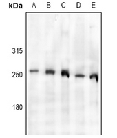
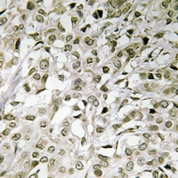
Application
| WB, IHC |
|---|---|
| Primary Accession | P21675 |
| Other Accession | Q80UV9 |
| Reactivity | Human, Mouse, Rat, Pig, Bovine, Dog |
| Host | Rabbit |
| Clonality | Polyclonal |
| Calculated MW | 214714 Da |
| Gene ID | 6872 |
|---|---|
| Other Names | BA2R; CCG1; CCGS; TAF2A; Transcription initiation factor TFIID subunit 1; Cell cycle gene 1 protein; TBP-associated factor 250 kDa; p250; Transcription initiation factor TFIID 250 kDa subunit; TAF(II)250; TAFII-250; TAFII250 |
| Target/Specificity | KLH-conjugated synthetic peptide encompassing a sequence within the center region of human TAF1. The exact sequence is proprietary. |
| Dilution | WB~~WB (1/500 - 1/1000), IH (1/50 - 1/200) IHC~~1:100~500 |
| Format | Liquid in 0.42% Potassium phosphate, 0.87% Sodium chloride, pH 7.3, 30% glycerol, and 0.09% (W/V) sodium azide. |
| Storage | Store at -20 °C.Stable for 12 months from date of receipt |
| Name | TAF1 (HGNC:11535) |
|---|---|
| Synonyms | BA2R, CCG1, CCGS, TAF2A |
| Function | The TFIID basal transcription factor complex plays a major role in the initiation of RNA polymerase II (Pol II)-dependent transcription (PubMed:33795473). TFIID recognizes and binds promoters with or without a TATA box via its subunit TBP, a TATA-box-binding protein, and promotes assembly of the pre-initiation complex (PIC) (PubMed:33795473). The TFIID complex consists of TBP and TBP-associated factors (TAFs), including TAF1, TAF2, TAF3, TAF4, TAF5, TAF6, TAF7, TAF8, TAF9, TAF10, TAF11, TAF12 and TAF13 (PubMed:33795473). TAF1 is the largest component and core scaffold of the TFIID complex, involved in nucleating complex assembly (PubMed:25412659, PubMed:27007846, PubMed:33795473). TAF1 forms a promoter DNA binding subcomplex of TFIID, together with TAF7 and TAF2 (PubMed:33795473). Contains novel N- and C-terminal Ser/Thr kinase domains which can autophosphorylate or transphosphorylate other transcription factors (PubMed:25412659, PubMed:8625415). Phosphorylates TP53 on 'Thr-55' which leads to MDM2- mediated degradation of TP53 (PubMed:25412659). Phosphorylates GTF2A1 and GTF2F1 on Ser residues (PubMed:25412659). Possesses DNA-binding activity (PubMed:25412659). Essential for progression of the G1 phase of the cell cycle (PubMed:11278496, PubMed:15053879, PubMed:2038334, PubMed:8450888, PubMed:8625415, PubMed:9660973, PubMed:9858607). Exhibits histone acetyltransferase activity towards histones H3 and H4 (PubMed:15870300). |
| Cellular Location | Nucleus |

Thousands of laboratories across the world have published research that depended on the performance of antibodies from Abcepta to advance their research. Check out links to articles that cite our products in major peer-reviewed journals, organized by research category.
info@abcepta.com, and receive a free "I Love Antibodies" mug.

Provided below are standard protocols that you may find useful for product applications.
Background
KLH-conjugated synthetic peptide encompassing a sequence within the center region of human TAF1. The exact sequence is proprietary.
If you have used an Abcepta product and would like to share how it has performed, please click on the "Submit Review" button and provide the requested information. Our staff will examine and post your review and contact you if needed.
If you have any additional inquiries please email technical services at tech@abcepta.com.
Ordering Information
Other Products
Shipping Information
