


 Foundational characteristics of cancer include proliferation, angiogenesis, migration, evasion of apoptosis, and cellular immortality. Find key markers for these cellular processes and antibodies to detect them.
 The SUMOplot™ Analysis Program predicts and scores sumoylation sites in your protein. SUMOylation is a post-translational modification involved in various cellular processes, such as nuclear-cytosolic transport, transcriptional regulation, apoptosis, protein stability, response to stress, and progression through the cell cycle.
 The Autophagy Receptor Motif Plotter predicts and scores autophagy receptor binding sites in your protein. Identifying proteins connected to this pathway is critical to understanding the role of autophagy in physiological as well as pathological processes such as development, differentiation, neurodegenerative diseases, stress, infection, and cancer.
Anti-ATP5D Antibody
Rabbit polyclonal antibody to ATP5D
- SPECIFICATION
- CITATIONS
- PROTOCOLS
- BACKGROUND

Application
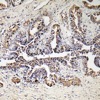
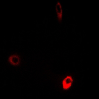
| WB, IF/IC, IHC |
|---|---|
| Primary Accession | P30049 |
| Other Accession | Q9D3D9 |
| Reactivity | Human, Mouse, Rat, Monkey, Bovine |
| Host | Rabbit |
| Clonality | Polyclonal |
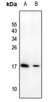
| Calculated MW | 17490 Da |
| Gene ID | 513 |
|---|---|
| Other Names | ATP synthase subunit delta mitochondrial; F-ATPase delta subunit |
| Target/Specificity | KLH-conjugated synthetic peptide encompassing a sequence within the center region of human ATP5D. The exact sequence is proprietary. |
| Dilution | WB~~WB (1/500 - 1/1000), IH (1/50 - 1/200), IF/IC (1/100 - 1/500) IF/IC~~N/A IHC~~1:100~500 |
| Format | Liquid in 0.42% Potassium phosphate, 0.87% Sodium chloride, pH 7.3, 30% glycerol, and 0.09% (W/V) sodium azide. |
| Storage | Store at -20 °C.Stable for 12 months from date of receipt |
| Name | ATP5F1D (HGNC:837) |
|---|---|
| Function | Subunit delta, of the mitochondrial membrane ATP synthase complex (F(1)F(0) ATP synthase or Complex V) that produces ATP from ADP in the presence of a proton gradient across the membrane which is generated by electron transport complexes of the respiratory chain (Probable) (PubMed:37244256). ATP synthase complex consist of a soluble F(1) head domain - the catalytic core - and a membrane F(1) domain - the membrane proton channel (PubMed:37244256). These two domains are linked by a central stalk rotating inside the F(1) region and a stationary peripheral stalk (PubMed:37244256). During catalysis, ATP synthesis in the catalytic domain of F(1) is coupled via a rotary mechanism of the central stalk subunits to proton translocation (Probable). In vivo, can only synthesize ATP although its ATP hydrolase activity can be activated artificially in vitro (By similarity). With the central stalk subunit gamma, is essential for the biogenesis of F(1) catalytic part of the ATP synthase complex namely in the formation of F1 assembly intermediate (PubMed:29499186). |
| Cellular Location | Mitochondrion. Mitochondrion inner membrane. |

Thousands of laboratories across the world have published research that depended on the performance of antibodies from Abcepta to advance their research. Check out links to articles that cite our products in major peer-reviewed journals, organized by research category.
info@abcepta.com, and receive a free "I Love Antibodies" mug.

Provided below are standard protocols that you may find useful for product applications.
Background
KLH-conjugated synthetic peptide encompassing a sequence within the center region of human ATP5D. The exact sequence is proprietary.
If you have used an Abcepta product and would like to share how it has performed, please click on the "Submit Review" button and provide the requested information. Our staff will examine and post your review and contact you if needed.
If you have any additional inquiries please email technical services at tech@abcepta.com.
Ordering Information
Other Products
Shipping Information
