


 Foundational characteristics of cancer include proliferation, angiogenesis, migration, evasion of apoptosis, and cellular immortality. Find key markers for these cellular processes and antibodies to detect them.
 The SUMOplot™ Analysis Program predicts and scores sumoylation sites in your protein. SUMOylation is a post-translational modification involved in various cellular processes, such as nuclear-cytosolic transport, transcriptional regulation, apoptosis, protein stability, response to stress, and progression through the cell cycle.
 The Autophagy Receptor Motif Plotter predicts and scores autophagy receptor binding sites in your protein. Identifying proteins connected to this pathway is critical to understanding the role of autophagy in physiological as well as pathological processes such as development, differentiation, neurodegenerative diseases, stress, infection, and cancer.
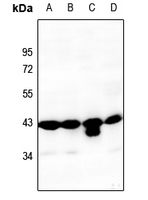
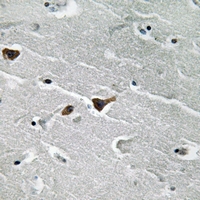
Anti-PAI1 Antibody
Rabbit polyclonal antibody to PAI1
- SPECIFICATION
- CITATIONS
- PROTOCOLS
- BACKGROUND

Application
| WB, IHC |
|---|---|
| Primary Accession | P05121 |
| Other Accession | P22777 |
| Reactivity | Human, Mouse, Rat |
| Host | Rabbit |
| Clonality | Polyclonal |
| Calculated MW | 45060 Da |
| Gene ID | 5054 |
|---|---|
| Other Names | PAI1; PLANH1; Plasminogen activator inhibitor 1; PAI; PAI-1; Endothelial plasminogen activator inhibitor; Serpin E1 |
| Target/Specificity | KLH-conjugated synthetic peptide encompassing a sequence within the center region of human PAI1. The exact sequence is proprietary. |
| Dilution | WB~~WB (1/500 - 1/1000), IH (1/50 - 1/200) IHC~~1:100~500 |
| Format | Liquid in 0.42% Potassium phosphate, 0.87% Sodium chloride, pH 7.3, 30% glycerol, and 0.09% (W/V) sodium azide. |
| Storage | Store at -20 °C.Stable for 12 months from date of receipt |
| Name | SERPINE1 |
|---|---|
| Synonyms | PAI1, PLANH1 |
| Function | Serine protease inhibitor. Inhibits TMPRSS7 (PubMed:15853774). Is a primary inhibitor of tissue-type plasminogen activator (PLAT) and urokinase-type plasminogen activator (PLAU). As PLAT inhibitor, it is required for fibrinolysis down-regulation and is responsible for the controlled degradation of blood clots (PubMed:17912461, PubMed:8481516, PubMed:9207454, PubMed:21925150). As PLAU inhibitor, it is involved in the regulation of cell adhesion and spreading (PubMed:9175705). Acts as a regulator of cell migration, independently of its role as protease inhibitor (PubMed:15001579, PubMed:9168821). It is required for stimulation of keratinocyte migration during cutaneous injury repair (PubMed:18386027). It is involved in cellular and replicative senescence (PubMed:16862142). Plays a role in alveolar type 2 cells senescence in the lung (By similarity). Is involved in the regulation of cementogenic differentiation of periodontal ligament stem cells, and regulates odontoblast differentiation and dentin formation during odontogenesis (PubMed:25808697, PubMed:27046084). |
| Cellular Location | Secreted. |
| Tissue Location | Expressed in endothelial cells (PubMed:2430793, PubMed:3097076). Found in plasma, platelets, and hepatoma and fibrosarcoma cells. |

Thousands of laboratories across the world have published research that depended on the performance of antibodies from Abcepta to advance their research. Check out links to articles that cite our products in major peer-reviewed journals, organized by research category.
info@abcepta.com, and receive a free "I Love Antibodies" mug.

Provided below are standard protocols that you may find useful for product applications.
Background
KLH-conjugated synthetic peptide encompassing a sequence within the center region of human PAI1. The exact sequence is proprietary.
If you have used an Abcepta product and would like to share how it has performed, please click on the "Submit Review" button and provide the requested information. Our staff will examine and post your review and contact you if needed.
If you have any additional inquiries please email technical services at tech@abcepta.com.
Ordering Information
Other Products
Shipping Information
