


 Foundational characteristics of cancer include proliferation, angiogenesis, migration, evasion of apoptosis, and cellular immortality. Find key markers for these cellular processes and antibodies to detect them.
 The SUMOplot™ Analysis Program predicts and scores sumoylation sites in your protein. SUMOylation is a post-translational modification involved in various cellular processes, such as nuclear-cytosolic transport, transcriptional regulation, apoptosis, protein stability, response to stress, and progression through the cell cycle.
 The Autophagy Receptor Motif Plotter predicts and scores autophagy receptor binding sites in your protein. Identifying proteins connected to this pathway is critical to understanding the role of autophagy in physiological as well as pathological processes such as development, differentiation, neurodegenerative diseases, stress, infection, and cancer.
Anti-PLC beta 3 Antibody
Rabbit polyclonal antibody to PLC beta 3
- SPECIFICATION
- CITATIONS: 1
- PROTOCOLS
- BACKGROUND

Application
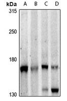
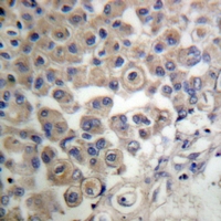
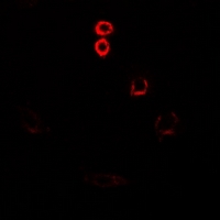
| WB, IF/IC, IHC |
|---|---|
| Primary Accession | Q01970 |
| Other Accession | P51432 |
| Reactivity | Human, Mouse, Rat |
| Host | Rabbit |
| Clonality | Polyclonal |
| Calculated MW | 138799 Da |
| Gene ID | 5331 |
|---|---|
| Other Names | 1-phosphatidylinositol 45-bisphosphate phosphodiesterase beta-3; Phosphoinositide phospholipase C-beta-3; Phospholipase C-beta-3; PLC-beta-3 |
| Target/Specificity | KLH-conjugated synthetic peptide encompassing a sequence within the center region of human PLC beta 3. The exact sequence is proprietary. |
| Dilution | WB~~WB (1/500 - 1/1000), IH (1/50 - 1/200), IF/IC (1/100 - 1/500) IF/IC~~N/A IHC~~1:100~500 |
| Format | Liquid in 0.42% Potassium phosphate, 0.87% Sodium chloride, pH 7.3, 30% glycerol, and 0.09% (W/V) sodium azide. |
| Storage | Store at -20 °C.Stable for 12 months from date of receipt |
| Name | PLCB3 {ECO:0000303|PubMed:20966218, ECO:0000312|EMBL:AAA77683.1} |
|---|---|
| Function | Catalyzes the production of the second messenger molecules diacylglycerol (DAG) and inositol 1,4,5-trisphosphate (IP3) (PubMed:20966218, PubMed:29122926, PubMed:37991948, PubMed:9188725). Key transducer of G protein-coupled receptor signaling: activated by G(q)/G(11) G alpha proteins downstream of G protein-coupled receptors activation (PubMed:20966218, PubMed:37991948). In neutrophils, participates in a phospholipase C-activating N-formyl peptide-activated GPCR (G protein-coupled receptor) signaling pathway by promoting RASGRP4 activation by DAG, to promote neutrophil functional responses (By similarity). |
| Cellular Location | Cytoplasm. Membrane {ECO:0000250|UniProtKB:Q99JE6}. Nucleus {ECO:0000250|UniProtKB:P51432} Note=And particulate fractions. {ECO:0000250|UniProtKB:Q99JE6} |
Research Areas
Citations ( 0 )

Application Protocols
Provided below are standard protocols that you may find useful for product applications.
Background
KLH-conjugated synthetic peptide encompassing a sequence within the center region of human PLC beta 3. The exact sequence is proprietary.
Abcepta welcomes feedback from its customers.
If you have used an Abcepta product and would like to share how it has performed, please click on the "Submit Review" button and provide the requested information. Our staff will examine and post your review and contact you if needed.
If you have any additional inquiries please email technical services at tech@abcepta.com.
$ 195.00
$ 385.00
Cat# AP61288
Ordering Information
United States
AlbaniaAustraliaAustriaBelgiumBosnia & HerzegovinaBrazilBulgariaCanadaCentral AmericaChinaCroatiaCyprusCzech RepublicDenmarkEstoniaFinlandFranceGermanyGreeceHong KongHungaryIcelandIndiaIndonesiaIrelandIsraelItalyJapanLatviaLithuaniaLuxembourgMacedoniaMalaysiaMaltaMexicoNetherlandsNew ZealandNorwayPakistanPolandPortugalRomaniaSerbiaSingaporeSlovakiaSloveniaSouth AfricaSouth KoreaSpainSwedenSwitzerlandTaiwanTurkeyUnited KingdomUnited StatesVietnamWorldwideOthers
USA Headquarters
(888) 735-7227 / (858) 622-0099 or (858) 875-1900
Other Products
Shipping Information
Domestic orders (in stock items)
Shipped out the same day. Orders placed after 1 PM (PST) will ship out the next business day.
International orders
Contact your local distributors
