


 Foundational characteristics of cancer include proliferation, angiogenesis, migration, evasion of apoptosis, and cellular immortality. Find key markers for these cellular processes and antibodies to detect them.
 The SUMOplot™ Analysis Program predicts and scores sumoylation sites in your protein. SUMOylation is a post-translational modification involved in various cellular processes, such as nuclear-cytosolic transport, transcriptional regulation, apoptosis, protein stability, response to stress, and progression through the cell cycle.
 The Autophagy Receptor Motif Plotter predicts and scores autophagy receptor binding sites in your protein. Identifying proteins connected to this pathway is critical to understanding the role of autophagy in physiological as well as pathological processes such as development, differentiation, neurodegenerative diseases, stress, infection, and cancer.
Anti-MID1 Antibody
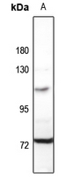
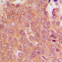
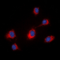
Rabbit polyclonal antibody to MID1
- SPECIFICATION
- CITATIONS
- PROTOCOLS
- BACKGROUND

Application
| WB, IF/IC, IHC |
|---|---|
| Primary Accession | O15344 |
| Other Accession | O70583 |
| Reactivity | Human, Mouse, Rat, Monkey, Dog |
| Host | Rabbit |
| Clonality | Polyclonal |
| Calculated MW | 75251 Da |
| Gene ID | 4281 |
|---|---|
| Other Names | FXY; RNF59; TRIM18; XPRF; E3 ubiquitin-protein ligase Midline-1; Midin; Putative transcription factor XPRF; RING finger protein 59; RING finger protein Midline-1; Tripartite motif-containing protein 18 |
| Target/Specificity | KLH-conjugated synthetic peptide encompassing a sequence within the N-term region of human MID1. The exact sequence is proprietary. |
| Dilution | WB~~WB (1/500 - 1/1000), IH (1/100 - 1/200), IF/IC (1/100 - 1/500) IF/IC~~N/A IHC~~1:100~500 |
| Format | Liquid in 0.42% Potassium phosphate, 0.87% Sodium chloride, pH 7.3, 30% glycerol, and 0.09% (W/V) sodium azide. |
| Storage | Store at -20 °C.Stable for 12 months from date of receipt |
| Name | MID1 |
|---|---|
| Synonyms | FXY, RNF59, TRIM18, XPRF |
| Function | Has E3 ubiquitin ligase activity towards IGBP1, promoting its monoubiquitination, which results in deprotection of the catalytic subunit of protein phosphatase PP2A, and its subsequent degradation by polyubiquitination. |
| Cellular Location | Cytoplasm. Cytoplasm, cytoskeleton. Cytoplasm, cytoskeleton, spindle. Note=Microtubule- associated. It is associated with microtubules throughout the cell cycle, co-localizing with cytoplasmic fibers in interphase and with the mitotic spindle and midbodies during mitosis and cytokinesis |
| Tissue Location | In the fetus, highest expression found in kidney, followed by brain and lung. Expressed at low levels in fetal liver. In the adult, most abundant in heart, placenta and brain |

Thousands of laboratories across the world have published research that depended on the performance of antibodies from Abcepta to advance their research. Check out links to articles that cite our products in major peer-reviewed journals, organized by research category.
info@abcepta.com, and receive a free "I Love Antibodies" mug.

Provided below are standard protocols that you may find useful for product applications.
Background
KLH-conjugated synthetic peptide encompassing a sequence within the N-term region of human MID1. The exact sequence is proprietary.
If you have used an Abcepta product and would like to share how it has performed, please click on the "Submit Review" button and provide the requested information. Our staff will examine and post your review and contact you if needed.
If you have any additional inquiries please email technical services at tech@abcepta.com.
Ordering Information
Other Products
Shipping Information
