


 Foundational characteristics of cancer include proliferation, angiogenesis, migration, evasion of apoptosis, and cellular immortality. Find key markers for these cellular processes and antibodies to detect them.
 The SUMOplot™ Analysis Program predicts and scores sumoylation sites in your protein. SUMOylation is a post-translational modification involved in various cellular processes, such as nuclear-cytosolic transport, transcriptional regulation, apoptosis, protein stability, response to stress, and progression through the cell cycle.
 The Autophagy Receptor Motif Plotter predicts and scores autophagy receptor binding sites in your protein. Identifying proteins connected to this pathway is critical to understanding the role of autophagy in physiological as well as pathological processes such as development, differentiation, neurodegenerative diseases, stress, infection, and cancer.
Anti-Perforin 1 Antibody
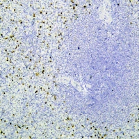
Mouse monoclonal antibody to Perforin 1
- SPECIFICATION
- CITATIONS
- PROTOCOLS
- BACKGROUND

Application
| IHC |
|---|---|
| Primary Accession | P14222 |
| Reactivity | Human |
| Host | Mouse |
| Clonality | Monoclonal |
| Calculated MW | 61377 Da |
| Gene ID | 5551 |
|---|---|
| Other Names | PFP; Perforin-1; P1; Cytolysin; Lymphocyte pore-forming protein; PFP |
| Target/Specificity | KLH-conjugated synthetic peptide encompassing a sequence within human Perforin 1. The exact sequence is proprietary. |
| Dilution | IHC~~1:100~500 |
| Format | Mouse IgG. Liquid in PBS containing 50% glycerol, 0.2% BSA and 0.09% (W/V) sodium azide. |
| Storage | Store at -20 °C.Stable for 12 months from date of receipt |
| Name | PRF1 |
|---|---|
| Synonyms | PFP |
| Function | Pore-forming protein that plays a key role in granzyme- mediated programmed cell death, and in defense against virus-infected or neoplastic cells (PubMed:20889983, PubMed:21037563, PubMed:24558045, PubMed:9058810, PubMed:9164947). Plays an important role in killing other cells that are recognized as non-self by the immune system, e.g. in transplant rejection or some forms of autoimmune disease (PubMed:9058810). Can insert into the membrane of target cells in its calcium-bound form, oligomerize and form large pores (PubMed:20889983, PubMed:21037563). Promotes cytolysis and apoptosis of target cells by mediating the passage and uptake of cytotoxic granzymes (PubMed:20038786, PubMed:20225066, PubMed:24558045, PubMed:32299851). Facilitates the delivery of cationic cargo protein, while anionic or neural proteins are not delivered efficiently (PubMed:24558045). Perforin pores allow the release of mature caspase-7 (CASP7) into the extracellular milieu (By similarity). |
| Cellular Location | Cytolytic granule. Secreted. Cell membrane; Multi-pass membrane protein. Endosome lumen. Note=Stored in cytolytic granules of cytolytic T-lymphocytes and secreted into the cleft between T- lymphocyte and target cell (PubMed:20038786). Inserts into the cell membrane of target cells and forms pores (PubMed:20889983). Membrane insertion and pore formation requires a major conformation change (PubMed:20889983). May be taken up via endocytosis involving clathrin- coated vesicles and accumulate in a first time in large early endosomes (PubMed:20038786). |

Thousands of laboratories across the world have published research that depended on the performance of antibodies from Abcepta to advance their research. Check out links to articles that cite our products in major peer-reviewed journals, organized by research category.
info@abcepta.com, and receive a free "I Love Antibodies" mug.

Provided below are standard protocols that you may find useful for product applications.
Background
KLH-conjugated synthetic peptide encompassing a sequence within human Perforin 1. The exact sequence is proprietary.
If you have used an Abcepta product and would like to share how it has performed, please click on the "Submit Review" button and provide the requested information. Our staff will examine and post your review and contact you if needed.
If you have any additional inquiries please email technical services at tech@abcepta.com.
Ordering Information
Other Products
Shipping Information
