


 Foundational characteristics of cancer include proliferation, angiogenesis, migration, evasion of apoptosis, and cellular immortality. Find key markers for these cellular processes and antibodies to detect them.
 The SUMOplot™ Analysis Program predicts and scores sumoylation sites in your protein. SUMOylation is a post-translational modification involved in various cellular processes, such as nuclear-cytosolic transport, transcriptional regulation, apoptosis, protein stability, response to stress, and progression through the cell cycle.
 The Autophagy Receptor Motif Plotter predicts and scores autophagy receptor binding sites in your protein. Identifying proteins connected to this pathway is critical to understanding the role of autophagy in physiological as well as pathological processes such as development, differentiation, neurodegenerative diseases, stress, infection, and cancer.
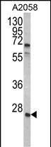
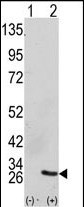
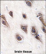
GRB2 Antibody (Center)
Purified Rabbit Polyclonal Antibody (Pab)
- SPECIFICATION
- CITATIONS
- PROTOCOLS
- BACKGROUND

Application
| WB, IHC-P, E |
|---|---|
| Primary Accession | P62993 |
| Other Accession | P62994, Q60631, Q6GPJ9, P87379 |
| Reactivity | Human |
| Predicted | Xenopus, Mouse, Rat |
| Host | Rabbit |
| Clonality | Polyclonal |
| Isotype | Rabbit IgG |
| Calculated MW | 25206 Da |
| Antigen Region | 89-118 aa |
| Gene ID | 2885 |
|---|---|
| Other Names | Growth factor receptor-bound protein 2, Adapter protein GRB2, Protein Ash, SH2/SH3 adapter GRB2, GRB2, ASH |
| Target/Specificity | This GRB2 antibody is generated from rabbits immunized with a KLH conjugated synthetic peptide between 89-118 amino acids from the Central region of human GRB2. |
| Dilution | WB~~1:1000 IHC-P~~1:50~100 E~~Use at an assay dependent concentration. |
| Format | Purified polyclonal antibody supplied in PBS with 0.09% (W/V) sodium azide. This antibody is prepared by Saturated Ammonium Sulfate (SAS) precipitation followed by dialysis against PBS. |
| Storage | Maintain refrigerated at 2-8°C for up to 2 weeks. For long term storage store at -20°C in small aliquots to prevent freeze-thaw cycles. |
| Precautions | GRB2 Antibody (Center) is for research use only and not for use in diagnostic or therapeutic procedures. |
| Name | GRB2 |
|---|---|
| Synonyms | ASH |
| Function | Non-enzymatic adapter protein that plays a pivotal role in precisely regulated signaling cascades from cell surface receptors to cellular responses, including signaling transduction and gene expression (PubMed:11726515, PubMed:37626338). Thus, participates in many biological processes including regulation of innate and adaptive immunity, autophagy, DNA repair or necroptosis (PubMed:35831301, PubMed:37626338, PubMed:38182563). Controls signaling complexes at the T-cell antigen receptor to facilitate the activation, differentiation, and function of T-cells (PubMed:36864087, PubMed:9489702). Mechanistically, engagement of the TCR leads to phosphorylation of the adapter protein LAT, which serves as docking site for GRB2 (PubMed:9489702). In turn, GRB2 establishes a a connection with SOS1 that acts as a guanine nucleotide exchange factor and serves as a critical regulator of KRAS/RAF1 leading to MAPKs translocation to the nucleus and activation (PubMed:12171928, PubMed:25870599). Functions also a role in B-cell activation by amplifying Ca(2+) mobilization and activation of the ERK MAP kinase pathway upon recruitment to the phosphorylated B-cell antigen receptor (BCR) (PubMed:25413232, PubMed:29523808). Plays a role in switching between autophagy and programmed necrosis upstream of EGFR by interacting with components of necrosomes including RIPK1 and with autophagy regulators SQSTM1 and BECN1 (PubMed:35831301, PubMed:38182563). Regulates miRNA biogenesis by forming a functional ternary complex with AGO2 and DICER1 (PubMed:37328606). Functions in the replication stress response by protecting DNA at stalled replication forks from MRE11-mediated degradation. Mechanistically, inhibits RAD51 ATPase activity to stabilize RAD51 on stalled replication forks (PubMed:38459011). Additionally, directly recruits and later releases MRE11 at DNA damage sites during the homology-directed repair (HDR) process (PubMed:34348893). |
| Cellular Location | Nucleus. Cytoplasm. Endosome. Golgi apparatus {ECO:0000250|UniProtKB:Q60631} |

Thousands of laboratories across the world have published research that depended on the performance of antibodies from Abcepta to advance their research. Check out links to articles that cite our products in major peer-reviewed journals, organized by research category.
info@abcepta.com, and receive a free "I Love Antibodies" mug.

Provided below are standard protocols that you may find useful for product applications.
Background
GRB2 binds the epidermal growth factor receptor and contains one SH2 domain and two SH3 domains. Its two SH3 domains direct complex formation with proline-rich regions of other proteins, and its SH2 domain binds tyrosine phosphorylated sequences.
References
Kondo,A., J. Biol. Chem. 283 (3), 1428-1436 (2008)
Morimatsu,M., Proc. Natl. Acad. Sci. U.S.A. 104 (46), 18013-18018 (2007)
Martinez,N., Cell. Signal. 19 (11), 2277-2285 (2007)
If you have used an Abcepta product and would like to share how it has performed, please click on the "Submit Review" button and provide the requested information. Our staff will examine and post your review and contact you if needed.
If you have any additional inquiries please email technical services at tech@abcepta.com.
Ordering Information
Other Products
Shipping Information
