


 Foundational characteristics of cancer include proliferation, angiogenesis, migration, evasion of apoptosis, and cellular immortality. Find key markers for these cellular processes and antibodies to detect them.
 The SUMOplot™ Analysis Program predicts and scores sumoylation sites in your protein. SUMOylation is a post-translational modification involved in various cellular processes, such as nuclear-cytosolic transport, transcriptional regulation, apoptosis, protein stability, response to stress, and progression through the cell cycle.
 The Autophagy Receptor Motif Plotter predicts and scores autophagy receptor binding sites in your protein. Identifying proteins connected to this pathway is critical to understanding the role of autophagy in physiological as well as pathological processes such as development, differentiation, neurodegenerative diseases, stress, infection, and cancer.
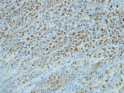
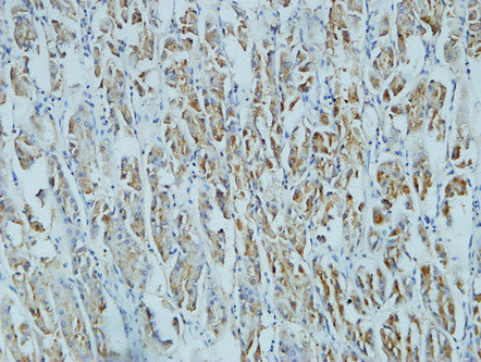
CD15 Monoclonal Antibody(Q89)
- SPECIFICATION
- CITATIONS
- PROTOCOLS
- BACKGROUND

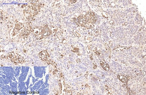
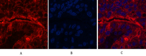
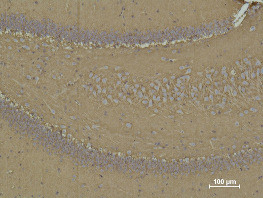
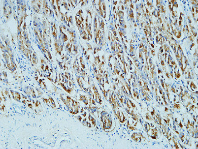
Application
| IHC-P, IF |
|---|---|
| Primary Accession | P22083 |
| Reactivity | Human |
| Host | Mouse |
| Clonality | Monoclonal |
| Calculated MW | 59084 Da |
| Gene ID | 2526 |
|---|---|
| Other Names | FUT4; ELFT; FCT3A; Alpha-(1, 3)-fucosyltransferase; ELAM-1 ligand fucosyltransferase; Fucosyltransferase 4; Fucosyltransferase IV; Fuc-TIV; FucT-IV; Galactoside 3-L-fucosyltransferase |
| Dilution | IHC-P~~N/A IF~~IHC 1:200 IF 1:50-200 |
| Format | PBS, pH 7.4, containing 0.09% (W/V) sodium azide as Preservative and 50% Glycerol. |
| Storage Conditions | -20℃ |
| Name | FUT4 {ECO:0000303|PubMed:29593094} |
|---|---|
| Function | [Isoform Short]: Catalyzes alpha(1->3) linkage of fucosyl moiety transferred from GDP-beta-L-fucose to N-acetyl glucosamine (GlcNAc) within type 2 lactosamine (LacNAc, Gal-beta(1->4)GlcNAc) glycan attached to N- or O-linked glycoproteins (PubMed:1702034, PubMed:1716630, PubMed:29593094). Robustly fucosylates nonsialylated distal LacNAc unit of the polylactosamine chain to form Lewis X antigen (CD15), a glycan determinant known to mediate important cellular functions in development and immunity. Fucosylates with lower efficiency sialylated LacNAc acceptors to form sialyl Lewis X and 6- sulfo sialyl Lewis X determinants that serve as recognition epitopes for C-type lectins (PubMed:1716630, PubMed:29593094). Together with FUT7 contributes to SELE, SELL and SELP selectin ligand biosynthesis and selectin-dependent lymphocyte homing, leukocyte migration and blood leukocyte homeostasis (By similarity). In a cell type specific manner, may also fucosylate the internal LacNAc unit of the polylactosamine chain to form VIM-2 antigen that serves as recognition epitope for SELE (PubMed:11278338, PubMed:1716630). |
| Cellular Location | Golgi apparatus, Golgi stack membrane; Single- pass type II membrane protein. Note=Membrane-bound form in trans cisternae of Golgi |
| Tissue Location | [Isoform Short]: Expressed at low levels in bone marrow-derived mesenchymal stem cells. |

Thousands of laboratories across the world have published research that depended on the performance of antibodies from Abcepta to advance their research. Check out links to articles that cite our products in major peer-reviewed journals, organized by research category.
info@abcepta.com, and receive a free "I Love Antibodies" mug.

Provided below are standard protocols that you may find useful for product applications.
Background
May catalyze alpha-1,3 glycosidic linkages involved in the expression of Lewis X/SSEA-1 and VIM-2 antigens.
If you have used an Abcepta product and would like to share how it has performed, please click on the "Submit Review" button and provide the requested information. Our staff will examine and post your review and contact you if needed.
If you have any additional inquiries please email technical services at tech@abcepta.com.
Ordering Information
Other Products
Shipping Information
