


 Foundational characteristics of cancer include proliferation, angiogenesis, migration, evasion of apoptosis, and cellular immortality. Find key markers for these cellular processes and antibodies to detect them.
 The SUMOplot™ Analysis Program predicts and scores sumoylation sites in your protein. SUMOylation is a post-translational modification involved in various cellular processes, such as nuclear-cytosolic transport, transcriptional regulation, apoptosis, protein stability, response to stress, and progression through the cell cycle.
 The Autophagy Receptor Motif Plotter predicts and scores autophagy receptor binding sites in your protein. Identifying proteins connected to this pathway is critical to understanding the role of autophagy in physiological as well as pathological processes such as development, differentiation, neurodegenerative diseases, stress, infection, and cancer.
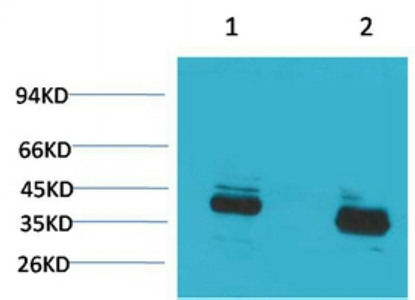
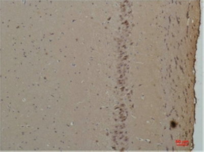
CABP Polyclonal Antibody
- SPECIFICATION
- CITATIONS
- PROTOCOLS
- BACKGROUND

Application
| WB, IHC-P |
|---|---|
| Primary Accession | Q9NZU7 |
| Reactivity | Rat, Mouse |
| Host | Rabbit |
| Clonality | Polyclonal |
| Calculated MW | 39838 Da |
| Gene ID | 9478 |
|---|---|
| Other Names | Calcium-binding protein 1 (CaBP1) (Calbrain) (Caldendrin) |
| Dilution | WB~~Western Blot: 1/500 - 1/2000. Immunohistochemistry: 1/100 - 1/300. ELISA: 1/40000. Not yet tested in other applications. IHC-P~~N/A |
| Format | Liquid in PBS containing 50% glycerol, 0.5% BSA and 0.09% (W/V) sodium azide. |
| Storage Conditions | -20℃ |
| Name | CABP1 |
|---|---|
| Function | Modulates calcium-dependent activity of inositol 1,4,5- triphosphate receptors (ITPRs) (PubMed:14570872). Inhibits agonist- induced intracellular calcium signaling (PubMed:15980432). Enhances inactivation and does not support calcium-dependent facilitation of voltage-dependent P/Q-type calcium channels (PubMed:11865310). Causes calcium-dependent facilitation and inhibits inactivation of L-type calcium channels by binding to the same sites as calmodulin in the C- terminal domain of CACNA1C, but has an opposite effect on channel function (PubMed:15140941). Suppresses the calcium-dependent inactivation of CACNA1D (By similarity). Inhibits TRPC5 channels (PubMed:15895247). Prevents NMDA receptor-induced cellular degeneration. Required for the normal transfer of light signals through the retina (By similarity). |
| Cellular Location | Cytoplasm, cytoskeleton. Cytoplasm, perinuclear region. Cell membrane; Lipid-anchor; Cytoplasmic side. Golgi apparatus Postsynaptic density. Note=L-CaBP1 is associated most likely with the cytoskeletal structures, whereas S-CaBP1 is localized at or near the plasma membrane. [Isoform S-CaBP1]: Cytoplasm, cell cortex. Cell membrane; Lipid-anchor Note=S-CaBP1 is localized at or near the plasma membrane |
| Tissue Location | Retina and brain. Somatodendritic compartment of neurons. Calbrain was found exclusively in brain where it is abundant in the hippocampus, habenular area in the epithalamus and in the cerebellum |

Thousands of laboratories across the world have published research that depended on the performance of antibodies from Abcepta to advance their research. Check out links to articles that cite our products in major peer-reviewed journals, organized by research category.
info@abcepta.com, and receive a free "I Love Antibodies" mug.

Provided below are standard protocols that you may find useful for product applications.
Background
Modulates calcium-dependent activity of inositol 1,4,5- triphosphate receptors (ITPRs)(PubMed:14570872). Inhibits agonist- induced intracellular calcium signaling (PubMed:15980432). Enhances inactivation and does not support calcium-dependent facilitation of voltage-dependent P/Q-type calcium channels (PubMed:11865310). Causes calcium-dependent facilitation and inhibits inactivation of L-type calcium channels by binding to the same sites as calmodulin in the C-terminal domain of CACNA1C, but has an opposite effect on channel function (PubMed:15140941). Suppresses the calcium-dependent inactivation of CACNA1D (By similarity). Inhibits TRPC5 channels (PubMed:15895247). Prevents NMDA receptor-induced cellular degeneration. Required for the normal transfer of light signals through the retina (By similarity).
If you have used an Abcepta product and would like to share how it has performed, please click on the "Submit Review" button and provide the requested information. Our staff will examine and post your review and contact you if needed.
If you have any additional inquiries please email technical services at tech@abcepta.com.
Ordering Information
Other Products
Shipping Information
