


 Foundational characteristics of cancer include proliferation, angiogenesis, migration, evasion of apoptosis, and cellular immortality. Find key markers for these cellular processes and antibodies to detect them.
 The SUMOplot™ Analysis Program predicts and scores sumoylation sites in your protein. SUMOylation is a post-translational modification involved in various cellular processes, such as nuclear-cytosolic transport, transcriptional regulation, apoptosis, protein stability, response to stress, and progression through the cell cycle.
 The Autophagy Receptor Motif Plotter predicts and scores autophagy receptor binding sites in your protein. Identifying proteins connected to this pathway is critical to understanding the role of autophagy in physiological as well as pathological processes such as development, differentiation, neurodegenerative diseases, stress, infection, and cancer.
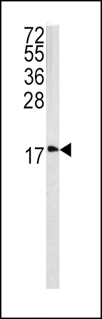
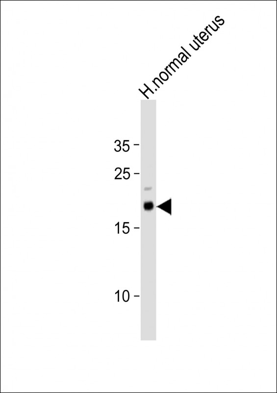
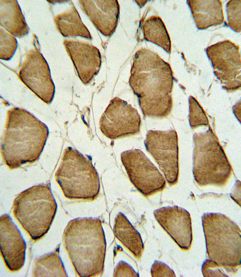
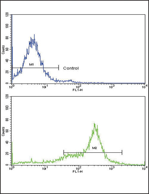
CAV3 Antibody (N-term)
Purified Rabbit Polyclonal Antibody (Pab)
- SPECIFICATION
- CITATIONS
- PROTOCOLS
- BACKGROUND

Application
| WB, IHC-P, FC, E |
|---|---|
| Primary Accession | P56539 |
| Other Accession | P51638, Q3ZDQ5, P51637, Q2KI43 |
| Reactivity | Human |
| Predicted | Bovine, Mouse, Pig, Rat |
| Host | Rabbit |
| Clonality | Polyclonal |
| Isotype | Rabbit IgG |
| Calculated MW | 17259 Da |
| Antigen Region | 5-31 aa |
| Gene ID | 859 |
|---|---|
| Other Names | Caveolin-3, M-caveolin, CAV3 |
| Target/Specificity | This CAV3 antibody is generated from rabbits immunized with a KLH conjugated synthetic peptide between 5-31 amino acids from the N-terminal region of human CAV3. |
| Dilution | WB~~1:500 IHC-P~~1:10~50 FC~~1:10~50 E~~Use at an assay dependent concentration. |
| Format | Purified polyclonal antibody supplied in PBS with 0.09% (W/V) sodium azide. This antibody is prepared by Saturated Ammonium Sulfate (SAS) precipitation followed by dialysis against PBS. |
| Storage | Maintain refrigerated at 2-8°C for up to 2 weeks. For long term storage store at -20°C in small aliquots to prevent freeze-thaw cycles. |
| Precautions | CAV3 Antibody (N-term) is for research use only and not for use in diagnostic or therapeutic procedures. |
| Name | CAV3 |
|---|---|
| Function | May act as a scaffolding protein within caveolar membranes. Interacts directly with G-protein alpha subunits and can functionally regulate their activity. May also regulate voltage-gated potassium channels. Plays a role in the sarcolemma repair mechanism of both skeletal muscle and cardiomyocytes that permits rapid resealing of membranes disrupted by mechanical stress (By similarity). Mediates the recruitment of CAVIN2 and CAVIN3 proteins to the caveolae (PubMed:19262564). |
| Cellular Location | Golgi apparatus membrane; Peripheral membrane protein. Cell membrane {ECO:0000250|UniProtKB:P51638}; Peripheral membrane protein. Membrane, caveola {ECO:0000250|UniProtKB:P51637}; Peripheral membrane protein. Cell membrane, sarcolemma {ECO:0000250|UniProtKB:P51637}. Note=Potential hairpin-like structure in the membrane. Membrane protein of caveolae (By similarity) |
| Tissue Location | Expressed predominantly in muscle. |

Thousands of laboratories across the world have published research that depended on the performance of antibodies from Abcepta to advance their research. Check out links to articles that cite our products in major peer-reviewed journals, organized by research category.
info@abcepta.com, and receive a free "I Love Antibodies" mug.

Provided below are standard protocols that you may find useful for product applications.
Background
CAV3 is a caveolin family member, which functions as a component of the caveolae plasma membranes found in most cell types. Caveolin proteins are proposed to be scaffolding proteins for organizing and concentrating certain caveolin-interacting molecules. Mutations identified in its gene lead to interference with protein oligomerization or intra-cellular routing, disrupting caveolae formation and resulting in Limb-Girdle muscular dystrophy type-1C (LGMD-1C), hyperCKemia or rippling muscle disease (RMD).
References
Garg,V., Biochem. Biophys. Res. Commun. 385 (3), 472-477 (2009)
Cai,C., . Biol. Chem. 284 (23), 15894-15902 (2009)
If you have used an Abcepta product and would like to share how it has performed, please click on the "Submit Review" button and provide the requested information. Our staff will examine and post your review and contact you if needed.
If you have any additional inquiries please email technical services at tech@abcepta.com.
Ordering Information
Other Products
Shipping Information
