


 Foundational characteristics of cancer include proliferation, angiogenesis, migration, evasion of apoptosis, and cellular immortality. Find key markers for these cellular processes and antibodies to detect them.
 The SUMOplot™ Analysis Program predicts and scores sumoylation sites in your protein. SUMOylation is a post-translational modification involved in various cellular processes, such as nuclear-cytosolic transport, transcriptional regulation, apoptosis, protein stability, response to stress, and progression through the cell cycle.
 The Autophagy Receptor Motif Plotter predicts and scores autophagy receptor binding sites in your protein. Identifying proteins connected to this pathway is critical to understanding the role of autophagy in physiological as well as pathological processes such as development, differentiation, neurodegenerative diseases, stress, infection, and cancer.
XPO1 Antibody (C-term)
Purified Rabbit Polyclonal Antibody (Pab)
- SPECIFICATION
- CITATIONS: 1
- PROTOCOLS
- BACKGROUND

Application
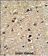
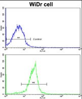
| WB, IHC-P, FC, E |
|---|---|
| Primary Accession | O14980 |
| Other Accession | Q80U96, Q6P5F9 |
| Reactivity | Human |
| Predicted | Mouse, Rat |
| Host | Rabbit |
| Clonality | Polyclonal |
| Isotype | Rabbit IgG |
| Calculated MW | 123386 Da |
| Antigen Region | 817-846 aa |
| Gene ID | 7514 |
|---|---|
| Other Names | Exportin-1, Exp1, Chromosome region maintenance 1 protein homolog, XPO1, CRM1 |
| Target/Specificity | This XPO1 antibody is generated from rabbits immunized with a KLH conjugated synthetic peptide between 817-846 amino acids from the C-terminal region of human XPO1. |
| Dilution | WB~~1:1000 IHC-P~~1:50~100 FC~~1:10~50 E~~Use at an assay dependent concentration. |
| Format | Purified polyclonal antibody supplied in PBS with 0.09% (W/V) sodium azide. This antibody is prepared by Saturated Ammonium Sulfate (SAS) precipitation followed by dialysis against PBS. |
| Storage | Maintain refrigerated at 2-8°C for up to 2 weeks. For long term storage store at -20°C in small aliquots to prevent freeze-thaw cycles. |
| Precautions | XPO1 Antibody (C-term) is for research use only and not for use in diagnostic or therapeutic procedures. |
| Name | XPO1 |
|---|---|
| Synonyms | CRM1 |
| Function | Mediates the nuclear export of cellular proteins (cargos) bearing a leucine-rich nuclear export signal (NES) and of RNAs. In the nucleus, in association with RANBP3, binds cooperatively to the NES on its target protein and to the GTPase RAN in its active GTP-bound form (Ran-GTP). Docking of this complex to the nuclear pore complex (NPC) is mediated through binding to nucleoporins. Upon transit of a nuclear export complex into the cytoplasm, disassembling of the complex and hydrolysis of Ran-GTP to Ran-GDP (induced by RANBP1 and RANGAP1, respectively) cause release of the cargo from the export receptor. The directionality of nuclear export is thought to be conferred by an asymmetric distribution of the GTP- and GDP-bound forms of Ran between the cytoplasm and nucleus. Involved in U3 snoRNA transport from Cajal bodies to nucleoli. Binds to late precursor U3 snoRNA bearing a TMG cap. |
| Cellular Location | Cytoplasm. Nucleus, nucleoplasm. Nucleus, Cajal body. Nucleus, nucleolus. Note=Located in the nucleoplasm, Cajal bodies and nucleoli. Shuttles between the nucleus/nucleolus and the cytoplasm |
| Tissue Location | Expressed in heart, brain, placenta, lung, liver, skeletal muscle, pancreas, spleen, thymus, prostate, testis, ovary, small intestine, colon and peripheral blood leukocytes. Not expressed in the kidney. |

Provided below are standard protocols that you may find useful for product applications.
Background
XPO1 mediates leucine-rich nuclear export signal (NES)-dependent protein transport. Exportin 1 specifically inhibits the nuclear export of Rev and U snRNAs. It is involved in the control of several cellular processes by controlling the localization of cyclin B, MPAK, and MAPKAP kinase 2. This protein also regulates NFAT and AP-1.
References
Shen,A., Neurosurgery 65 (1), 153-159 (2009)
Dong,X., Nat. Struct. Mol. Biol. 16 (5), 558-560 (2009)
If you have used an Abcepta product and would like to share how it has performed, please click on the "Submit Review" button and provide the requested information. Our staff will examine and post your review and contact you if needed.
If you have any additional inquiries please email technical services at tech@abcepta.com.
Ordering Information
Other Products
Shipping Information
