


 Foundational characteristics of cancer include proliferation, angiogenesis, migration, evasion of apoptosis, and cellular immortality. Find key markers for these cellular processes and antibodies to detect them.
 The SUMOplot™ Analysis Program predicts and scores sumoylation sites in your protein. SUMOylation is a post-translational modification involved in various cellular processes, such as nuclear-cytosolic transport, transcriptional regulation, apoptosis, protein stability, response to stress, and progression through the cell cycle.
 The Autophagy Receptor Motif Plotter predicts and scores autophagy receptor binding sites in your protein. Identifying proteins connected to this pathway is critical to understanding the role of autophagy in physiological as well as pathological processes such as development, differentiation, neurodegenerative diseases, stress, infection, and cancer.
MTM1 Antibody (C-term)
Purified Rabbit Polyclonal Antibody (Pab)
- SPECIFICATION
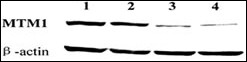
- CITATIONS: 1
- PROTOCOLS
- BACKGROUND

Application
| WB, E |
|---|---|
| Primary Accession | Q13496 |
| Reactivity | Human, Mouse |
| Host | Rabbit |
| Clonality | Polyclonal |
| Isotype | Rabbit IgG |
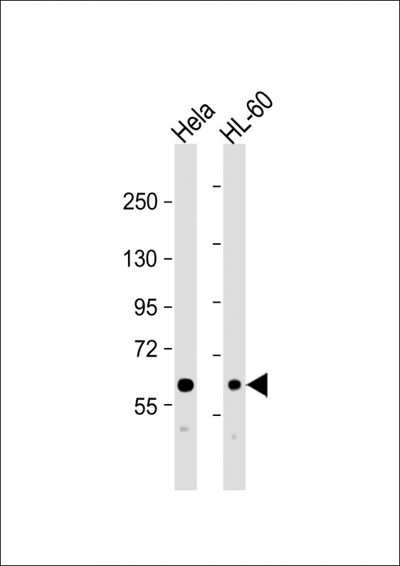
| Calculated MW | 69932 Da |
| Antigen Region | 566-594 aa |
| Gene ID | 4534 |
|---|---|
| Other Names | Myotubularin, Phosphatidylinositol-3, 5-bisphosphate 3-phosphatase, Phosphatidylinositol-3-phosphate phosphatase, MTM1, CG2 |
| Target/Specificity | This MTM1 antibody is generated from rabbits immunized with a KLH conjugated synthetic peptide between 566-594 amino acids from the C-terminal region of human MTM1. |
| Dilution | WB~~1:2000 E~~Use at an assay dependent concentration. |
| Format | Purified polyclonal antibody supplied in PBS with 0.09% (W/V) sodium azide. This antibody is prepared by Saturated Ammonium Sulfate (SAS) precipitation followed by dialysis against PBS. |
| Storage | Maintain refrigerated at 2-8°C for up to 2 weeks. For long term storage store at -20°C in small aliquots to prevent freeze-thaw cycles. |
| Precautions | MTM1 Antibody (C-term) is for research use only and not for use in diagnostic or therapeutic procedures. |
| Name | MTM1 (HGNC:7448) |
|---|---|
| Synonyms | CG2 |
| Function | Lipid phosphatase which dephosphorylates phosphatidylinositol 3-monophosphate (PI3P) and phosphatidylinositol 3,5-bisphosphate (PI(3,5)P2) (PubMed:10900271, PubMed:11001925, PubMed:12646134, PubMed:14722070). Has also been shown to dephosphorylate phosphotyrosine- and phosphoserine-containing peptides (PubMed:9537414). Negatively regulates EGFR degradation through regulation of EGFR trafficking from the late endosome to the lysosome (PubMed:14722070). Plays a role in vacuolar formation and morphology. Regulates desmin intermediate filament assembly and architecture (PubMed:21135508). Plays a role in mitochondrial morphology and positioning (PubMed:21135508). Required for skeletal muscle maintenance but not for myogenesis (PubMed:21135508). In skeletal muscles, stabilizes MTMR12 protein levels (PubMed:23818870). |
| Cellular Location | Cytoplasm. Cell membrane; Peripheral membrane protein. Cell projection, filopodium. Cell projection, ruffle. Late endosome. Cytoplasm, myofibril, sarcomere {ECO:0000250|UniProtKB:Q9Z2C5}. Note=Localizes as a dense cytoplasmic network (PubMed:11001925). Also localizes to the plasma membrane, including plasma membrane extensions such as filopodia and ruffles (PubMed:12118066). Predominantly located in the cytoplasm following interaction with MTMR12 (PubMed:12847286). Recruited to the late endosome following EGF stimulation (PubMed:14722070). In skeletal muscles, co-localizes with MTMR12 in the sarcomere (By similarity) {ECO:0000250|UniProtKB:Q9Z2C5, ECO:0000269|PubMed:11001925, ECO:0000269|PubMed:12118066, ECO:0000269|PubMed:12847286, ECO:0000269|PubMed:14722070} |

Provided below are standard protocols that you may find useful for product applications.
Background
MTM1 is a member of a protein family that encodes tyrosine phosphatases. Myotubularin is required for muscle cell differentiation and mutations in MTM1 have been identified as being responsible for X-linked myotubular myopathy. MTM1 is a potent phosphatidylinositol 3-phosphate phosphatase (PI(3)P). Mutations in the MTM1 gene that cause human myotubular myopathy dramatically reduce the ability of the phosphatase to dephosphorylate PI(3)P. The findings provided evidence that myotubularin exerts its effects during myogenesis by regulating the cellular levels of the inositol lipid PI(3)P.
References
Nandurkar, H.H., et al., Proc. Natl. Acad. Sci. U.S.A. 100(15):8660-8665 (2003).
Biancalana, V., et al., Hum. Genet. 112(2):135-142 (2003).
Wishart, M.J., et al., Trends Cell Biol. 12(12):579-585 (2002).
Herman, G.E., et al., Hum. Mutat. 19(2):114-121 (2002).
Sutton, I.J., et al., Neurology 57(5):900-902 (2001).
If you have used an Abcepta product and would like to share how it has performed, please click on the "Submit Review" button and provide the requested information. Our staff will examine and post your review and contact you if needed.
If you have any additional inquiries please email technical services at tech@abcepta.com.
Ordering Information
Other Products
Shipping Information
