


 Foundational characteristics of cancer include proliferation, angiogenesis, migration, evasion of apoptosis, and cellular immortality. Find key markers for these cellular processes and antibodies to detect them.
 The SUMOplot™ Analysis Program predicts and scores sumoylation sites in your protein. SUMOylation is a post-translational modification involved in various cellular processes, such as nuclear-cytosolic transport, transcriptional regulation, apoptosis, protein stability, response to stress, and progression through the cell cycle.
 The Autophagy Receptor Motif Plotter predicts and scores autophagy receptor binding sites in your protein. Identifying proteins connected to this pathway is critical to understanding the role of autophagy in physiological as well as pathological processes such as development, differentiation, neurodegenerative diseases, stress, infection, and cancer.
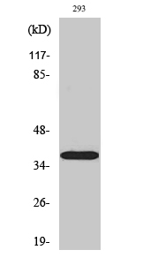
17β-HSD11 Polyclonal Antibody
- SPECIFICATION
- CITATIONS
- PROTOCOLS
- BACKGROUND

Application
| WB |
|---|---|
| Primary Accession | Q8NBQ5 |
| Reactivity | Human |
| Host | Rabbit |
| Clonality | Polyclonal |
| Calculated MW | 32964 Da |
| Gene ID | 51170 |
|---|---|
| Other Names | HSD17B11; DHRS8; PAN1B; PSEC0029; Estradiol 17-beta-dehydrogenase 11; 17-beta-hydroxysteroid dehydrogenase 11; 17-beta-HSD 11; 17bHSD11; 17betaHSD11; 17-beta-hydroxysteroid dehydrogenase XI; 17-beta-HSD XI; 17betaHSDXI; Cutaneous T-cell lym |
| Dilution | WB~~Western Blot: 1/500 - 1/2000. ELISA: 1/5000. Not yet tested in other applications. |
| Format | Liquid in PBS containing 50% glycerol, 0.5% BSA and 0.09% (W/V) sodium azide. |
| Storage Conditions | -20℃ |
| Name | HSD17B11 |
|---|---|
| Synonyms | DHRS8, PAN1B, SDR16C2 |
| Function | Can convert androstan-3-alpha,17-beta-diol (3-alpha-diol) to androsterone in vitro, suggesting that it may participate in androgen metabolism during steroidogenesis. May act by metabolizing compounds that stimulate steroid synthesis and/or by generating metabolites that inhibit it. Has no activity toward DHEA (dehydroepiandrosterone), or A- dione (4-androste-3,17-dione), and only a slight activity toward testosterone to A-dione. Tumor-associated antigen in cutaneous T-cell lymphoma. |
| Cellular Location | Endoplasmic reticulum {ECO:0000250|UniProtKB:Q9EQ06}. Lipid droplet {ECO:0000250|UniProtKB:Q9EQ06}. Note=Redistributed from the endoplasmic reticulum to lipids droplets in the cell upon induction of lipids droplet formation. {ECO:0000250|UniProtKB:Q9EQ06} |
| Tissue Location | Present at high level in steroidogenic cells such as syncytiotrophoblasts, sebaceous gland, Leydig cells, and granulosa cells of the dominant follicle and corpus luteum. In lung, it is detected in the ciliated epithelium and in acini of adult trachea, in bronchioles, but not in alveoli. In the eye, it is detected in the nonpigmented epithelium of the ciliary body and, at lower level, in the inner nuclear layer of the retina (at protein level). Widely expressed Highly expressed in retina, pancreas, kidney, liver, lung, adrenal, small intestine, ovary and heart. |

Thousands of laboratories across the world have published research that depended on the performance of antibodies from Abcepta to advance their research. Check out links to articles that cite our products in major peer-reviewed journals, organized by research category.
info@abcepta.com, and receive a free "I Love Antibodies" mug.

Provided below are standard protocols that you may find useful for product applications.
Background
Can convert androstan-3-alpha,17-beta-diol (3-alpha- diol) to androsterone in vitro, suggesting that it may participate in androgen metabolism during steroidogenesis. May act by metabolizing compounds that stimulate steroid synthesis and/or by generating metabolites that inhibit it. Has no activity toward DHEA (dehydroepiandrosterone), or A-dione (4-androste-3,17-dione), and only a slight activity toward testosterone to A-dione. Tumor- associated antigen in cutaneous T-cell lymphoma.
If you have used an Abcepta product and would like to share how it has performed, please click on the "Submit Review" button and provide the requested information. Our staff will examine and post your review and contact you if needed.
If you have any additional inquiries please email technical services at tech@abcepta.com.
Ordering Information
Shipping Information
