


 Foundational characteristics of cancer include proliferation, angiogenesis, migration, evasion of apoptosis, and cellular immortality. Find key markers for these cellular processes and antibodies to detect them.
 The SUMOplot™ Analysis Program predicts and scores sumoylation sites in your protein. SUMOylation is a post-translational modification involved in various cellular processes, such as nuclear-cytosolic transport, transcriptional regulation, apoptosis, protein stability, response to stress, and progression through the cell cycle.
 The Autophagy Receptor Motif Plotter predicts and scores autophagy receptor binding sites in your protein. Identifying proteins connected to this pathway is critical to understanding the role of autophagy in physiological as well as pathological processes such as development, differentiation, neurodegenerative diseases, stress, infection, and cancer.
AChRα10 Polyclonal Antibody
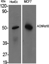
.jpg)
- SPECIFICATION
- CITATIONS
- PROTOCOLS
- BACKGROUND

Application
| WB |
|---|---|
| Primary Accession | Q9GZZ6 |
| Reactivity | Human, Mouse, Rat |
| Host | Rabbit |
| Clonality | Polyclonal |
| Calculated MW | 49705 Da |
| Gene ID | 57053 |
|---|---|
| Other Names | CHRNA10; NACHRA10; Neuronal acetylcholine receptor subunit alpha-10; Nicotinic acetylcholine receptor subunit alpha-10; NACHR alpha-10 |
| Dilution | WB~~Western Blot: 1/500 - 1/2000. ELISA: 1/20000. Not yet tested in other applications. |
| Format | Liquid in PBS containing 50% glycerol, 0.5% BSA and 0.09% (W/V) sodium azide. |
| Storage Conditions | -20℃ |
| Name | CHRNA10 (HGNC:13800) |
|---|---|
| Synonyms | NACHRA10 |
| Function | Component of neuronal acetylcholine receptors (nAChRs) that function as pentameric, ligand-gated cation channels with high calcium permeability. nAChRs are excitatory neurotrasnmitter receptors formed by a collection of nAChR subunits. Each nAchR subunit confers differential attributes to channel properties, including activation, deactivation and desensitization kinetics, pH sensitivity, cation permeability, and binding to allosteric modulators (Probable). Forms heteropentamers with CHRNA9. Expressed in the inner ear, in sympathetic neurons and in other non-neuronal cells, such as skin keratinocytes and lymphocytes (PubMed:11752216, PubMed:15531379). nAChR formed by CHRNA9:CHRNA10 is involved in modulation of auditory stimuli. The channel is permeable to a range of divalent cations including calcium, the influx of which may activate a potassium current which hyperpolarizes the cell membrane. In the ear, mediates synaptic transmission between efferent olivocochlear fibers and hair cells of the cochlea, this may lead to a reduction in basilar membrane motion, altering the activity of auditory nerve fibers and reducing the range of dynamic hearing (PubMed:11752216). This may protect against acoustic trauma. May also regulate keratinocyte adhesion (By similarity). |
| Cellular Location | Synaptic cell membrane {ECO:0000250|UniProtKB:Q9JLB5}; Multi-pass membrane protein. Cell membrane {ECO:0000250|UniProtKB:Q9JLB5}; Multi-pass membrane protein |
| Tissue Location | Expressed in inner-ear tissue, tonsil, immortalized B-cells, cultured T-cells and peripheral blood lymphocytes |

Thousands of laboratories across the world have published research that depended on the performance of antibodies from Abcepta to advance their research. Check out links to articles that cite our products in major peer-reviewed journals, organized by research category.
info@abcepta.com, and receive a free "I Love Antibodies" mug.

Provided below are standard protocols that you may find useful for product applications.
Background
Ionotropic receptor with a probable role in the modulation of auditory stimuli. Agonist binding may induce an extensive change in conformation that affects all subunits and leads to opening of an ion-conducting channel across the plasma membrane. The channel is permeable to a range of divalent cations including calcium, the influx of which may activate a potassium current which hyperpolarizes the cell membrane. In the ear, this may lead to a reduction in basilar membrane motion, altering the activity of auditory nerve fibers and reducing the range of dynamic hearing. This may protect against acoustic trauma.
If you have used an Abcepta product and would like to share how it has performed, please click on the "Submit Review" button and provide the requested information. Our staff will examine and post your review and contact you if needed.
If you have any additional inquiries please email technical services at tech@abcepta.com.
Ordering Information
Other Products
Shipping Information
