


 Foundational characteristics of cancer include proliferation, angiogenesis, migration, evasion of apoptosis, and cellular immortality. Find key markers for these cellular processes and antibodies to detect them.
 The SUMOplot™ Analysis Program predicts and scores sumoylation sites in your protein. SUMOylation is a post-translational modification involved in various cellular processes, such as nuclear-cytosolic transport, transcriptional regulation, apoptosis, protein stability, response to stress, and progression through the cell cycle.
 The Autophagy Receptor Motif Plotter predicts and scores autophagy receptor binding sites in your protein. Identifying proteins connected to this pathway is critical to understanding the role of autophagy in physiological as well as pathological processes such as development, differentiation, neurodegenerative diseases, stress, infection, and cancer.
PIK4CB Antibody (Center)
Purified Rabbit Polyclonal Antibody (Pab)
- SPECIFICATION
- CITATIONS
- PROTOCOLS
- BACKGROUND

Application
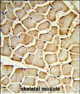
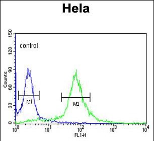
| FC, IHC-P, WB, E |
|---|---|
| Primary Accession | Q9UBF8 |
| Other Accession | O08561, Q8BKC8, Q49GP3, O02810 |
| Reactivity | Human |
| Predicted | Bovine, Zebrafish, Mouse, Rat |
| Host | Rabbit |
| Clonality | Polyclonal |
| Isotype | Rabbit IgG |
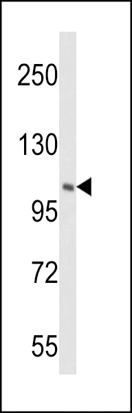
| Calculated MW | 91379 Da |
| Antigen Region | 519-548 aa |
| Gene ID | 5298 |
|---|---|
| Other Names | Phosphatidylinositol 4-kinase beta, PI4K-beta, PI4Kbeta, PtdIns 4-kinase beta, NPIK, PI4K92, PI4KB, PIK4CB |
| Target/Specificity | This PIK4CB antibody is generated from rabbits immunized with a KLH conjugated synthetic peptide between 519-548 amino acids from the Central region of human PIK4CB. |
| Dilution | FC~~1:10~50 IHC-P~~1:50~100 WB~~1:1000 E~~Use at an assay dependent concentration. |
| Format | Purified polyclonal antibody supplied in PBS with 0.09% (W/V) sodium azide. This antibody is prepared by Saturated Ammonium Sulfate (SAS) precipitation followed by dialysis against PBS. |
| Storage | Maintain refrigerated at 2-8°C for up to 2 weeks. For long term storage store at -20°C in small aliquots to prevent freeze-thaw cycles. |
| Precautions | PIK4CB Antibody (Center) is for research use only and not for use in diagnostic or therapeutic procedures. |
| Name | PI4KB (HGNC:8984) |
|---|---|
| Synonyms | PIK4CB |
| Function | Phosphorylates phosphatidylinositol (PI) in the first committed step in the production of the second messenger inositol- 1,4,5,-trisphosphate (PIP). May regulate Golgi disintegration/reorganization during mitosis, possibly via its phosphorylation. Involved in Golgi-to-plasma membrane trafficking (By similarity) (PubMed:10559940, PubMed:11277933, PubMed:12749687, PubMed:9405935). May play an important role in the inner ear development. |
| Cellular Location | Endomembrane system. Mitochondrion outer membrane; Peripheral membrane protein. Rough endoplasmic reticulum membrane; Peripheral membrane protein. Golgi apparatus. Golgi apparatus membrane. Cytoplasm, perinuclear region. Note=Found in the outer membrane of mitochondria and membranes of the rough endoplasmic reticulum. Recruited to the Golgi complex by the small GTPase ARF to stimulate the synthesis of phosphatidylinositol 4,5- bisphosphate (PIP2) on the Golgi complex. Recruited to the Golgi apparatus membrane by ACBD3 (PubMed:24672044, PubMed:27009356, PubMed:28289207). GGA2 is also involved in the recruitment (PubMed:28289207). |
| Tissue Location | Widely expressed with highest levels in heart, skeletal muscle, pancreas, testis and ovary. Weakly expressed in liver (PubMed:9020160, PubMed:9405935, PubMed:9405938). Expressed in the innear ear in the epithelium of the spinal organ of corti |

Thousands of laboratories across the world have published research that depended on the performance of antibodies from Abcepta to advance their research. Check out links to articles that cite our products in major peer-reviewed journals, organized by research category.
info@abcepta.com, and receive a free "I Love Antibodies" mug.

Provided below are standard protocols that you may find useful for product applications.
Background
PIK4CB phosphorylates phosphatidylinositol (PI) in the first committed step in the production of the second messenger inositol-1,4,5,-trisphosphate (PIP). It may regulate Golgi disintegration/reorganization during mitosis, possibly via its phosphorylation.
References
Jeganathan,S., et.al., Mol. Cell. Biol. 28 (14), 4549-4561 (2008) Pizarro-Cerda,J., et.al., Cell. Microbiol. 9 (10), 2381-2390 (2007)
If you have used an Abcepta product and would like to share how it has performed, please click on the "Submit Review" button and provide the requested information. Our staff will examine and post your review and contact you if needed.
If you have any additional inquiries please email technical services at tech@abcepta.com.
Ordering Information
Other Products
Shipping Information
